
Page 15 - DT 13-1 March 2023
P. 15
14-19-Vinayak article-Q8_6-7-8-Ivoclar.qxd 3/27/2023 1:47 PM Page 2
multidisciplinary section 15
DENTAL TECHNOLOGY, JANUARY-MARCH 2023
STAGE II - STAGED OCCLUSAL REHABILITATION
After pre alignment was completed, following chairside
deprogramming, fresh diagnostic impressions, Facebow
transfer and photos were taken and sent to the laboratory.
(Precision Dental Studio, Mumbai) 1
With the help of extra oral and intra oral photographs,
a 2D Smile Simulation was done adhering to Golden
Proportion. This help us verify the size, shape and position
of teeth.
Upon agreement of Size, Shape & Morphology of the
design file, the next step was to print a diagnostic. 2
The laboratory was asked to fabricate and send 3D
designed and printed models with raised vertical dimen-
sion by 2-3mm. A putty index of the 3D printed models
were made using addition silicone (Honigum, DMG). The
patient’s teeth were spot etched (Ultraetch, ULTRADENT)
and spot bonded (Scotchbond, 3M ) and the 3D designed
raised vertical and restored teeth were transferred into the
patients’ mouth using APT technique (Aesthetic
Provisional Temporary) The raised bite was equilibrated
bilaterally to remove any interferences with 200 micron
occlusal marking paper (Bausch). The patient was left to
test drive the raised vertical for 3 weeks and monitored for
para joint related discomfort. 1
After thorough evaluation and stabilization we decided
to prep the teeth and shift the patient to milled lab made
CAD CAM interim PMMA crown with the same vertical. 1
The printed PMMA provisionals were printed in Next
Dent 3D System. This system provides a Bio Compatible
resin that can be used as a long term provisional, are aes-
thetic in nature and has better strength compared to con-
ventional provisionals. 2
STAGE III: STAGED OCCLUSAL REHABILITATION WITH PMMA
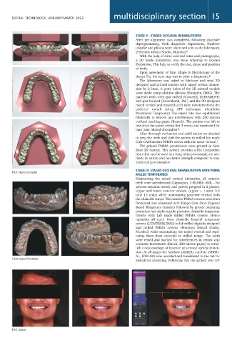
FIG 3: Aligner simulation
MILLED TEMPORARIES
Maintaining the raised vertical dimension, all anterior
teeth were anesthetized (Lignospan, 1:80,000) ADR – No
adverse reaction noted) and grossly prepped in 2 phases.
Upper and lower anterior sextant (upper + Lower 3-3
total 12 units) while maintaining posterior vertical with
the chairside temps. The anterior PMMA crowns were then
fabricated and cemented with Tempo Cem (Non Eugenol
Based Temporary Cement) followed by grossly preparing
posteriors and replacing the posterior chairside temporary
crowns with Lab made milled PMMA crowns. Hence
replacing all teeth from chairside bisacryl temporary
crowns (LUXATEMP, DMG) to lab milled digitally designed
and milled PMMA crowns (Precision Dental Studio,
Mumbai) while maintaining the raised vertical and repli-
cating them from chairside to milled temps. The teeth
were tested and marked for interferences in centric and
eccentric movements (Baush, 200 micron paper) to estab-
lish a new envelope of function at a raised vertical dimen-
sion. At all stages the facebow (ADLER) and bite (VIRTU-
AL, IVOCAR) was recorded and transferred to the lab for
FIG 4: Aligner Print Model
articulator mounting. Following this the patient was left
FIG 5: 2D DSD