
Page 26 - Dental Practice August 2022
P. 26
26-30-Richard-Q8:18-22-Lanka Mahesh.qxd 8/17/2022 6:13 PM Page 1
implantology section
COMPLEX PROBLEMS SOLVED
WITH BIOLOGICAL MINDSET
RICHARD WINTER
This is a case study of how an implant centered treatment plan
resulted in a non- surgical solution for a patient with pain, missing
teeth, and severely mal-posed dentition with mobility.
DYNAMIC TREATMENT SOLUTIONS
Dentists are specialists in diagnosing and treating problems of the
stomatognathic system. Typically, weare presented with problems
that can be defined as a subset of an anatomic issue such as a tooth
ache resulting from pulpitis or a cracked tooth. This problem-
focused diagnosis is the “bread and butter” of general dental prac-
tice. When examining a patient, we are taught to examine for oral-
cancer, occlusal problems, periodontal or endodontic issues, caries,
etc. but when multi-factorial problems exist, we must creatively
present plans that will address disease processes while honouring a
patient’s chief complaints, concerns and financial limits.
This case study highlights issues that we are faced with daily FIG 1: Full face pre-operatively
which involves when to extract teeth and place implants, how long
to treat periodontally challenged teeth and how to improve esthetic
difficulties in a patient with severe occlusal disharmonies.
CHIEF COMPLAINTS
This patient presented with a chief complaint of pain when chewing
and dissatisfaction with her smile. In examining her dentition, it was
noted that she had a lateral incisor in lingual version, Class II mal-
occlusion with a deep bite that resulted in her biting her incisive
papillae with normal chewing, and a severely canted occlusal plane
and severe fremitus with localized severe AAPIV Periodontitis.
In discussing her treatment, he expressed the desire to “pull her
teeth and give her implants.” There was Class III mobility of #6, 10,
12, 23-26, and several posterior molars. FIG 2: Relaxed smile photo
Her full-face smile photo revealsa large 3mm diastema with a
rotated canine and a slight grimace in maximum intercuspation
position (Figure 1). In relaxed smile position, it is noted that the
lower anteriors are in contact with her incisive papillae (Figure 2).
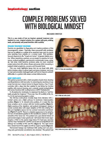
Slight opening of her dentition and occlusal photograph reveals a
canted occlusal plane with super eruption of #22 into missing #11
sites and highlights reasons for her occlusal traumatism and fremi-
tus (Figures 3 and 4). There is a stippled appearance to her gingiva,
but loss of interdental papillae in the mandibular anterior is repre-
sentative of her bone loss. The patient had a past dental history of
periodontal surgery 4 years previously. Left lateral view shows super
eruption of #22 to the crestal ridge where #11 had been removed
and the “step” between #21 and #22 can be seen to be significant and
etiology for mobility of #10 and #12 (Figure 5). The right lateral
view reveals that #10 is angled and #6 has super-erupted (Figure 6).
Periodontal therapy was instituted with scaling and root plan-
ning and a periodontal consultation was obtained. The patient had FIG 3: Retracted photo slight disclusion
26 Dental Practice // July-August 2022 // Vol 18 No 4