
Page 13 - DP Vol 21 No1_Neat
P. 13
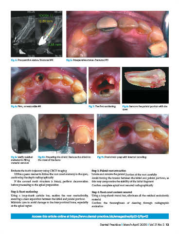
Fig 4: Preoperative status: fractured #11  Fig 5: Preoperative status: fractured #11


           Fig 6: Firm, unrestorable #11                        Fig 7: The first sectioning  Fig 8: Remove the palatal portion with the
                                                                                       apex


           Fig 9: Verify residual   Fig 10: Preparing the shield. Reduce the shield to   Fig 11: Final shield prep with internal bevelling
           endodontic filling   the crest of the bone
           material removal


           Evaluate the tooth trajectory using CBCT imaging.      Step 3: Palatal root extraction
              Utilize a peeso reamer to follow the root canal anatomy to the apex,   Luxate and remove the palatal portion of the root carefully.
           confirming the depth radiographically.                 Avoid forcing the luxator between the labial and palatal portions, as
              If the  coronal tooth structure is intact, perform decoronation   this may compromise the stability of the labial fragment.
           before proceeding to the apical preparation.           Confirm complete apical root removal radiographically.

           Step 2: Root sectioning                                Step 4: Root canal content removal
           Using a long-shank carbide bur, section the root mesiodistally,   Using a long-shank round bur, eliminate all the residual endodontic
           ensuring a clean separation between the labial and palatal portions.  material.
           Maintain care to avoid damage to the interproximal bone, especially   Confirm the  thoroughness of cleaning through  radiographic
           at the apical region.                                  evaluation.


                          Access this article online at https://www.dental-practice.biz/emagazine/dp21-2/#p=12

                                                                        Dental Practice I March-April 2025 I Vol 21 No 2  13