
Page 62 - DP Vol 21 No1_Neat
P. 62
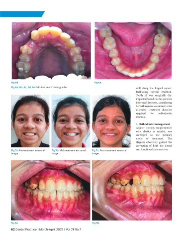
Fig 6d                                                  Fig 6e
           Fig 6a, 6b, 6c, 6d, 6e: Mid-treatment photographs                                   wall along the lingual aspect,
                                                                                               facilitating normal eruption.
                                                                                               Tooth 13 was surgically dis-
                                                                                               impacted based on the patient’s
                                                                                               informed decision, considering
                                                                                               her willingness to commit to the
                                                                                               extended treatment duration
                                                                                               required  for  orthodontic
                                                                                               traction.

                                                                                               3. Orthodontic management
                                                                                               Aligner therapy, supplemented
                                                                                               with elastics as needed, was
                                                                                               employed as the primary
                                                                                               mode   of  treatment.  The
                                                                                               aligners effectively guided the
                                                                                               correction of both the dental
           Fig 7a: Pre-treatment extraoral   Fig 7b: Mid-treatment extraoral   Fig 7c: Post-treatment extraoral   and functional asymmetries.
           image                      image                      image


           Fig 8a                                                 Fig 8b
           62 Dental Practice I March-April 2025 I Vol 21 No 2