
Page 57 - DP Vol 21 No1_Neat
P. 57
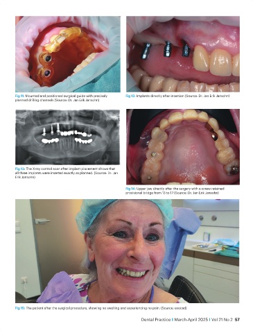
Fig 11: Mounted and positioned surgical guide with precisely   Fig 12: Implants directly after insertion (Source: Dr. Jan Erik Jansohn)
           planned drilling channels (Source: Dr. Jan Erik Jansohn)


           Fig 13: The X-ray control scan after implant placement shows that
           all three implants were inserted exactly as planned. (Source: Dr. Jan
           Erik Jansohn)


                                                                Fig 14: Upper jaw directly after the surgery with a screw-retained
                                                                provisional bridge from 13 to 17 (Source: Dr. Jan Erik Jansohn)


           Fig 15: The patient after the surgical procedure, showing no swelling and experiencing no pain. (Source: exocad)

                                                                        Dental Practice I March-April 2025 I Vol 21 No 2  57