
Page 33 - DP Vol 22 No 1
P. 33
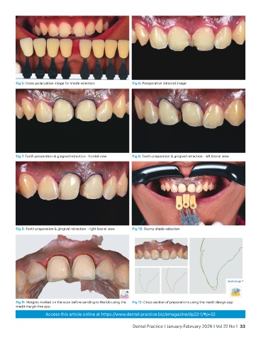
Fig 5: Cross polarization image for shade selection    Fig 6: Preoperative intraoral image


           Fig 7: Tooth preparation & gingival retraction - frontal view  Fig 8: Tooth preparation & gingival retraction - left lateral view


           Fig 9: Tooth preparation & gingival retraction - right lateral view  Fig 10: Stump shade selection


           Fig 11: Margins marked on the scan before sending to the lab using the   Fig 12: Cross section of preparations using the medit design app
           medit margin line app
                         Access this article online at https://www.dental-practice.biz/emagazine/dp22-1/#p=32

                                                                  Dental Practice I January-February 2026 I Vol 22 No 1  33