
Page 40 - DP Vol 22 No 1
P. 40
IMPLANTOLOGY
IMPLANT PLACEMENT AND SIMULTANEOUS GBR
Yazad Gandhi
THE SITUATION
A young male in his 30’s desired a fixed rehabilitation for
the second quadrant following removal of a failing FPD.
History revealed a surgical extraction of the maxillary
third molar in the same quadrant a few years ago.
Implant placement was carried out with a prosthetic
stent, hard tissue augmentation was done via direct sinus-
lift at the same stage using Geistlich Bio-Oss® large particle
with autogenous cortical shavings from the native site. The
grafted site was covered using a native collagen barrier,
Geistlich Bio-Gide®.
The quadrant was followed up for a period of 6 months
before commencing the prosthetic phase.
THE CHALLENGES
A large perforation of the Schneiderian membrane at the
disto-inferior angle of the osteotomy.
Fig 1: Pre-op OPG
THE APPROACH
Implant osteotomies were completed. The perforation was
sealed using collagen fleece followed by a second layer of
native collagen barrier Geistlich Bio-Gide®. Following this,
the biomaterial mix (Geistlich Bio-Oss® large particle with
autogenous cortical shavings) was inserted carefully to
ensure complete delivery to the medial aspect.
Fixtures were driven to the desired depth.
The window was covered using a second native collagen
barrier, Geistlich Bio-Gide®.
THE OUTCOME
At 6 months optimal regeneration was achieved, and the
fixtures were ready for loading.
CONCLUSION
Sinus augmentation is a predictable procedure to build
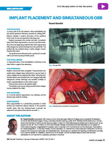
vertical and horizontal osseous volume in the posterior Fig 2: Guided pins are placed in the position
maxilla given that the evidence-based protocols are
adhered to and optimal biomaterials are used. n
ABOUT THE AUTHOR
Dr. Yazad Gandhi graduated with honours from King George’s Medical College and completed his Master’s
in Oral & Maxillofacial Surgery there, including implant training under Prof. Wilfried Schilli (Germany). He has
advanced training in hard and soft tissue surgery under Prof. Karl Kahnberg (Sweden) and in endoscopic sinus
surgery in the UK. He is a Fellow of the International College of Dentists and the International Congress of
Oral Implantologists, a registered ITI speaker, and a member of ITI Switzerland and the European Association
for Osseointegration. A Ginwala Oration Award recipient, Dr. Gandhi has conducted and mentored numerous
implant CDE programs nationally and internationally. He serves as a reviewer for leading maxillofacial journals,
is an opinion leader for Geistlich, BioHorizons and Versah. He has multiple national and international
publications and maintains a specialty practice in Mumbai.
40 Dental Practice I January-February 2026 I Vol 22 No 1 contd. on page 42