Page 12 - DT Vol 13 No 2
        P. 12
     10-15-Mike Prosperino-Q8_6-7-8-Ivoclar.qxd  6/19/2023  7:02 PM  Page 3
              12 cosmetic section                                                                  DENTAL TECHNOLOGY, APRIL-JUNE 2023
             F FIG 16 6                            F FIG 17 7                                      F FIG 18 8
                                                    G
               1
                                                    I
                                                                                                      1
                                                                                                    G
              I
                                                                                                    I
              G
                                                     1
                                                                                                      2
              G
                                                     2
                                                                                                    I
                                                                                                    G
               1
                                                    G
                                                    I
              I
             F FIG 19 9                            F FIG 20 0                                      F FIG 21 1
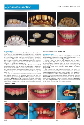
             CLINICAL CASE 2                                                required for rehabilitation (Figure 28).
             A 17-year-old patient presented to the office because she was dissat-
             isfied with her smile, particularly the shape and colour of her cen-  LABORATORY STEPS
             tral incisors. On intraoral clinical examination, there was evidence  We created a plaster model with the white GC Fujirock extra-hard
             of a class II malocclusion with deep bite, marked tooth crowding,  plaster after receiving the impressions and cleaning them with ultra-
             and fracture of the upper central incisors. Specifically, the patient  sonic and disinfection treatments (Figure 29).
             had suffered severe blunt trauma following an accidental fall, dur-  Following  that,  we  applied  the  platinum  foil  to  plaster
             ing  which  the  upper  central  incisors  chipped  in  the  mesial  area  models  (Figure  30).  One  of  the  first  and  most  essential  tasks
             involving the incisal angle (Figure 22).                       in  the  layering  in  cases  like  this  is  to  restrict  the  flow  of  light
               The  malocclusion  was  corrected  after  about  2  years  of  fixed  using  high-opacity  ceramics  that  prevent  the  excessive  path  of
             orthodontic therapy. Still, the patient was unsatisfied with the aes-  light  in  the  incisal  zones,  precisely  where  damage  is  evident
             thetic result, and the defect due to the previous fracture of the cen-  (Figure 31).
             tral incisors remained. A team with the dental technician then pro-  After  layering  the  work  with  polychromatic  Vita  Lumex  AC
             ceeded to study the case and to take impressions for wax-up and  ceramic masses, we proceeded for model analysis and the correct
             mock-up.                                                       shapes for the rehabilitation with golden powders (Yeti Dental) for
               An aesthetic-periodontal evaluation revealed the need to correct  the correct form and surface texture (Figure 32). Next, the ceram-
             the  gingival  architecture  to  harmonise  the  smile.  We  used  the  ics  are  glazed  and  polished  using  abrasive  rubbers  and  polishers
             detailed wax-up and mockup (Figure 23) to perform  mucogingi-  (Figure 33). Finally, the ceramics are extracted from the platinum
             val surgery (Figures 24 to 27). Next, with the dental technician,  sheet. (Note the delicate structure of ceramic veneers) (Figures 34
             we decided to conduct an aesthetic-functional rehabilitation using  and 35). In Figures 36 to 38, we can see the ceramics cemented
             ideal feldspar ceramic veneers with the minimal dental preparations  in the mouth about 15 days after their cementation.
             CLINICAL CASE 2
              G
                                                                                                    G
                                                                                                    I
              I
             F FIG 22 2                            F FIG 23 3                                      F FIG 24 4
                                                                                                      2
                                                    I
               2
                                                    G
                                                     2
                                                                                                   2
               2
                                                      I
             F FIG 25 5                              F FIG 26 6                                  F FIG 27 7
              I
              G
                                                                                                 I
                                                                                                  G
                                                      G
                                                        2


