


Page 18 - Dental Practice South Asia 18-6
P. 18
multidisciplinary section


              THE DIGITAL APPROACH IN THE MANAGEMENT OF


                 ANTERIOR SURGICAL-PROSTHETIC TREATMENT


                                                      ROBERTO MOLINARI, MD


              With  this  article  I  want  to  propose  a  new
              approach  to  digital  dentistry,  showing  new
              surgical prosthetic work protocols in terms of
              complex aesthetic rehabilitations in the ante-
              rior, with particular reference to patients who
              exhibit a “Gummy Smile”.
                More precisely, I would like to introduce
              the digital work protocol I adopt when I need
              to preserve the original morphology of soft
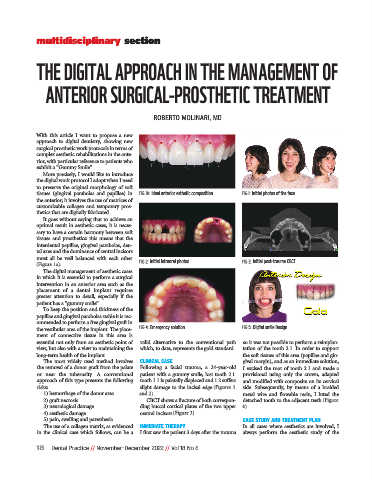
              tissues  (gingival  parabolas  and  papillae)  in  FIG 1A: Ideal anterior esthetic composition   FIG 1: Initial photos of the face
              the anterior; it involves the use of matrices of
              customizable  collagen  and  temporary  pros-
              thetics that are digitally fabricated.
                It goes without saying that to achieve an
              optimal result in aesthetic cases, it is neces-
              sary to have a certain harmony between soft
              tissues  and  prosthetics:  this  means  that  the
              interdental papillae, gingival parabolas, den-
              tal axes and the dominance of central incisors
              must  all  be  well  balanced  with  each  other
              (Figure 1a).                       FIG 2: Initial intraoral photos     FIG 3: Initial post-trauma CBCT
                The digital management of aesthetic cases
              in which it is essential to perform a surgical
              intervention in an anterior area such as the
              placement  of  a  dental  implant  requires
              greater  attention  to  detail,  especially  if  the
              patient has a “gummy smile”.
                To keep the position and thickness of the
              papillae and gingival parabolas stable it is rec-
              ommended to perform a free gingival graft in
              the vestibular area of the implant. The place-  FIG 4: Emergency solution   FIG 5: Digital smile Design
              ment  of  connective  tissue  in  this  area  is
              essential not only from an aesthetic point of  valid  alternative  to  the  conventional  path  so it was not possible to perform a reimplan-
              view, but also with a view to maintaining the  which, to date, represents the gold standard.  tation of the tooth 2.1. In order to support
              long-term health of the implant.                                       the soft tissues of this area (papillae and gin-
                The  most  widely  used  method  involves  CLINICAL CASE             gival margin), and as an immediate solution,
              the removal of a donor graft from the palate  Following  a  facial  trauma,  a  24-year-old  I excised the root of tooth 2.1 and made a
              or  near  the  tuberosity.  A  conventional  patient with a gummy smile, lost tooth 2.1.  provisional  using  only  the  crown,  adapted
              approach of this type presents the following  tooth 1.1 is palatally displaced and 1.2 suffers  and modified with composite on its cervical
              risks:                             slight damage to the incisal edge  (Figures 1  side.  Subsequently,  by  means  of  a  braided
                1) hemorrhage of the donor area   and 2).                            metal  wire  and  flowable  resin,  I  luted  the
                2) graft necrosis                   CBCT shows a fracture of both correspon-  detached tooth to the adjacent teeth (Figure
                3) neurological damage           ding buccal cortical plates of the two upper  4).
                4) aesthetic damage              central incisors (Figure 3).
                5) pain, swelling and paresthesia                                    CASE STUDY AND TREATMENT PLAN
                The use of a collagen matrix, as evidenced  IMMEDIATE THERAPY        In  all  cases  where  aesthetics  are  involved,  I
              in  the  clinical  case  which  follows,  can  be  a  I first saw the patient 3 days after the trauma  always  perform  the  aesthetic  study  of  the

              18   Dental Practice // November-December 2022 // Vol 18 No 6

