


Page 22 - Dental Practice South Asia 18-6
P. 22
multidisciplinary section


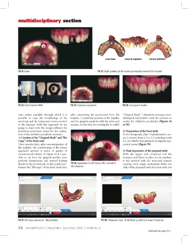
             FIG 31: Suture                                         FIG 32: Digital planning of the second provisional screwed to the implant


             FIG 33: Milled tooth in PMMA        FIG 34: Provisional in position     FIG 35: Healing at 3 months

              ware  makes  available,  through  which  it  is  after  unscrewing  the  provisional  from  the  “Gingival Mask”, I shared the necessary mor-
              possible  to  copy  the  morphology  of  the  implant, I copied the position of the papillae  phological information with the software to
              mock-up and the temporary crown screwed  and the gingival parabola with the intra-oral  realize the definitive prosthetics (Figures 36
              to  the  implant.  With  this  approach  we  are  scanner. In this way, by creating the so-called  to 38).
              going to share with the design software the
              anatomies  previously  tested  for  the  realiza-                      2) Preparation of the front teeth
              tion of the definitive prosthetic ceramics.                            In the therapeutic plan I had planned to cre-
              1) Creation of the “Gingival Mask” and “Bio                            ate 5 veneers, from 1.3 to 2.3 excluding tooth
              Copy” of the front teeth                                               2.1, for which I had planned an implant sup-
              Three months later, after osseointegration of                          ported crown (Figure 39).
              the implant, the conditioning of the tissues
              appeared  optimal  in  terms  of  quality  of                          3) Final impression of the prepared teeth
              reconstructed volume. In Figure 36 it is pos-                          With  the  upper  arch  prepared  and  the
              sible  to  see  how  the  gingival  profiles  were                     Scanpost and Tbase in place on the implant,
              perfectly  harmonious  and  natural  looking                           it  was  marked  with  the  intra-oral  scanner
              thanks to the provisional. At this point I per-  FIG 36: Appearance of soft tissues after removal of  copying  every  single  anatomical  detail  not
              formed the "Biocopy" of the front teeth and,  the temporary            only of the prepared teeth but even teeth not


             FIG 37: Soft tissue impression “Gingival Mask”           FIG 38: “Biogeneric Copy” of the Mock-up and the Screwed Provisional

              22   Dental Practice // November-December 2022 // Vol 18 No 6
                                                                                                       Continued on page 24 >>

