
Page 26 - DP.qxd
P. 26
prosthetic section
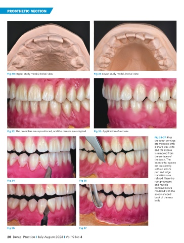
Fig 30: Upper study model, incisal view Fig 31: Lower study model, incisal view
Fig 32: The premolars are repositioned, and the canines are adapted Fig 33: Application of red wax
Fig 34-37: First
the tooth cervixes
are modeled with
a sharp wax knife
and the excess
is removed from
the surfaces of
the teeth. The
interdental spaces
are cut cleanly
with an artist’s
pen and edge
transitions are
refined. Then the
Fig 34 Fig 35 root processes
and muscle
concavities are
modeled with the
spoon-shaped
back of the wax
knife
Fig 36 Fig 37
26 Dental Practice i July-August 2023 i Vol 19 No 4