


Page 44 - DP Vol 20 No 3
P. 44
OrthOdOntic  sectiOn


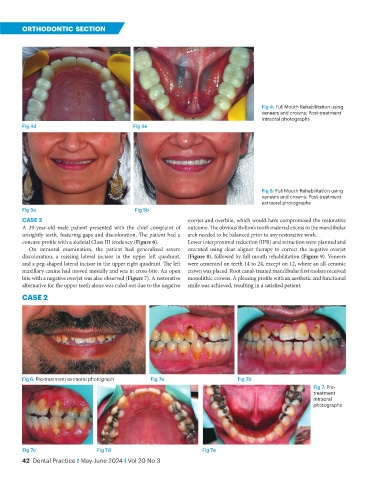
                                                                                           Fig 4: Full Mouth Rehabilitation using
                                                                                           veneers and crowns: Post-treatment
                                                                                           intraoral photographs
           Fig 4d                               Fig 4e


                                                                                           Fig 5: Full Mouth Rehabilitation using
                                                                                           veneers and crowns: Post-treatment
                                                                                           extraoral photographs
           Fig 5a                                Fig 5b
           CASE 2                                                 overjet and overbite, which would have compromised the restorative
           A 39-year-old male patient presented with the chief complaint of   outcome. The obvious Bolton’s tooth material excess in the mandibular
           unsightly teeth, featuring gaps and discoloration. The patient had a   arch needed to be balanced prior to any restorative work.
           concave profile with a skeletal Class III tendency (Figure 6).   Lower interproximal reduction (IPR) and retraction were planned and
              On intraoral examination, the patient had generalized severe   executed using clear aligner therapy to correct the negative overjet
           discoloration, a missing lateral incisor in the upper left quadrant,   (Figure 8), followed by full mouth rehabilitation (Figure 9). Veneers
           and a peg-shaped lateral incisor in the upper right quadrant. The left   were cemented on teeth 14 to 24, except on 12, where an all-ceramic
           maxillary canine had moved mesially and was in cross-bite. An open   crown was placed. Root canal-treated mandibular first molars received
           bite with a negative overjet was also observed (Figure 7). A restorative   monolithic crowns. A pleasing profile with an aesthetic and functional
           alternative for the upper teeth alone was ruled out due to the negative   smile was achieved, resulting in a satisfied patient.
           cAse 2


           Fig 6: Pre-treatment extraoral photograph  Fig 7a                       Fig 7b
                                                                                                            Fig 7: Pre-
                                                                                                            treatment
                                                                                                            Intraoral
                                                                                                            photographs


           Fig 7c                   Fig 7d                             Fig 7e
           42  Dental Practice I May-June 2024 I Vol 20 No 3

