
Page 19 - DP Vol 21 No1_Neat
P. 19
goals, irrespective of the treatment approach, are the achievement of Secondary treatment modalities for malocclusion or malunion
stable occlusion and normal mandibular function. Studies by August secondary to condylar fractures include occlusal splint therapy,
et al., Ylikontiola et al., and Parton et al. have reported an age-related physical therapy, selective grinding equilibration, orthodontic, and
increase in postoperative neurosensory disturbances. 7,8,9 Additionally, prosthetic correction.¹³ Surgical treatments such as arthroplasty, ramus
Peacock et al. found that patients older than 40 had longer hospital osteotomy, and joint replacement with TMJ prosthesis come with their
stays and a higher likelihood of hardware removal compared to own risks and costs. Disadvantages of alloplastic joint replacement
younger patients.¹⁰ Kriwalsky et al. also demonstrated that older include higher cost and hardware failure.¹⁴ All patients undergoing gap
patients are more prone to poor outcomes, such as inadequate fracture arthroplasty or joint reconstruction run the risk of potential injury to
healing, when managed surgically.¹¹ the facial nerve, Frey syndrome, and parotid gland injury.
Once satisfactory TMJ function is restored, post-traumatic The patient had come to believe that she must “live with” the
malocclusion can be addressed through either conservative or anterior and right-side posterior open bite, since the only treatment
surgical interventions. The duration of time between trauma and suggested by her previous dental surgeon was TMJ surgery—without
correction plays a significant role in determining the appropriate any guarantee of outcomes.
treatment. Mild post-traumatic malocclusions in patients with The patient was pleasantly surprised to learn that her request to
healthy teeth and periodontium can be treated conservatively through replace her crowns would also provide the significant additional
occlusal equilibration, prosthetic reconstruction, or orthodontics.¹² benefit of restoring her occlusion to a nearly normal level through
These conservative methods, which focus on modifying the shape prosthetic rehabilitation—a plan that had never been presented to her
and position of the teeth, can effectively correct post-traumatic by her previous dentists.
malocclusions without introducing significant risks.
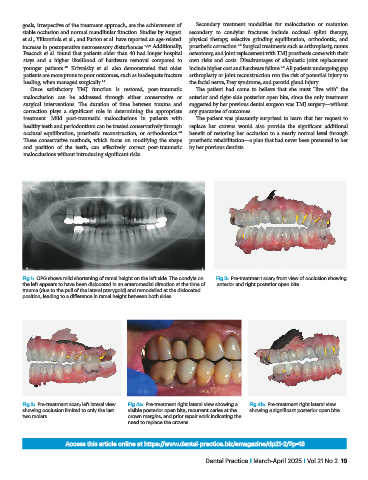
Fig 1: OPG shows mild shortening of ramal height on the left side. The condyle on Fig 2: Pre-treatment scan; front view of occlusion showing
the left appears to have been dislocated in an anteromedial direction at the time of anterior and right posterior open bite.
trauma (due to the pull of the lateral pterygoid) and remodelled at the dislocated
position, leading to a difference in ramal height between both sides.
Fig 3: Pre-treatment scan; left lateral view Fig 4a: Pre-treatment right lateral view showing a Fig 4b: Pre-treatment right lateral view
showing occlusion limited to only the last visible posterior open bite, recurrent caries at the showing a significant posterior open bite.
two molars. crown margins, and prior repair work indicating the
need to replace the crowns.
Access this article online at https://www.dental-practice.biz/emagazine/dp21-2/#p=18
Dental Practice I March-April 2025 I Vol 21 No 2 19