
Page 20 - DP Vol 21 No1_Neat
P. 20
PROSTHETICS
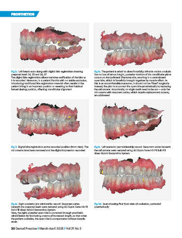
Fig 5: Left lateral scan along with digital bite registration showing Fig 6: The patient is asked to close forcefully; left-side molars occlude.
prepared teeth 24, 25 and 36, 37. Due to loss of ramus height, posterior rotation of the mandibular plane
The digital bite registration allows real-time verification of the bite as occurs on the ipsilateral (fracture) side, resulting in a contralateral
it is recorded. Moreover, in a patient like this with no stable occlusion, open bite, which is forcefully brought together by muscle effort. As
introducing traditional bite registration material often results in the this is an uncomfortable maneuver, it should not be “fixed” surgically.
patient biting in an incorrect position or reverting to their habitual Instead, the plan is to correct the open bite prosthetically by replacing
forced closing position, affecting mandibular alignment. the old crowns. Importantly, no virgin teeth need to be cut — only the
old crowns with recurrent caries, which require replacement anyway,
are addressed.
Fig 7: Digital bite registration at the recorded position (front view). The Fig 8: Left posterior jaw relationship record. Recurrent caries beneath
old crowns have been removed and the digital impression recorded. the old crowns were restored using GC Equia Forte HD Fil Bulk Fill
Glass Hybrid Restorative System.
Fig 9: Right posterior jaw relationship record. Recurrent caries Fig 10: Scan showing final front view of occlusion, corrected
beneath the prepared teeth were restored using GC Equia Forte HD Fil prosthetically.
Bulk Fill Glass Hybrid Restorative System.
Here, the right posterior open bite is corrected through prosthetic
rehabilitation by fabricating crowns of increased length, so that when
the patient occludes, the open bite is compensated without muscle
strain.
20 Dental Practice I March-April 2025 I Vol 21 No 2