
Page 21 - DP Vol 21 No1_Neat
P. 21
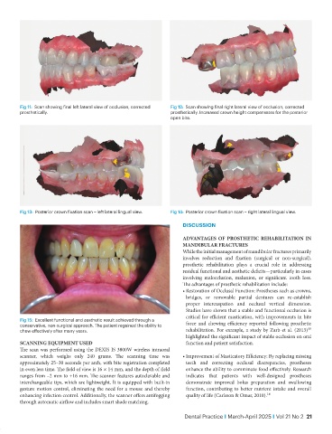
Fig 11:  Scan showing final left lateral view of occlusion, corrected   Fig 12:  Scan showing final right lateral view of occlusion, corrected
           prosthetically.                                         prosthetically. Increased crown height compensates for the posterior
                                                                   open bite.


           Fig 13:  Posterior crown fixation scan – left lateral lingual view.  Fig 14:  Posterior crown fixation scan – right lateral lingual view.

                                                                       DISCUSSION

                                                                       ADVANTAGES OF PROSTHETIC REHABILITATION IN
                                                                       MANDIBULAR FRACTURES
                                                                       While the initial management of mandibular fractures primarily
                                                                       involves reduction and fixation (surgical or non-surgical),
                                                                       prosthetic rehabilitation plays a crucial role in addressing
                                                                       residual functional and aesthetic deficits—particularly in cases
                                                                       involving malocclusion, malunion, or significant tooth loss.
                                                                       The advantages of prosthetic rehabilitation include:
                                                                       •  Restoration of Occlusal Function: Prostheses such as crowns,
                                                                         bridges, or removable partial dentures can re-establish
                                                                         proper intercuspation and occlusal vertical dimension.
                                                                         Studies have shown that a stable and functional occlusion is
                                                                         critical for efficient mastication, with improvements in bite
           Fig 15:  Excellent functional and aesthetic result achieved through a
           conservative, non-surgical approach. The patient regained the ability to   force  and  chewing  efficiency  reported  following  prosthetic
           chew effectively after many years.                            rehabilitation. For example, a study by Zarb et al. (2013)¹⁵
                                                                         highlighted the significant impact of stable occlusion on oral
           SCANNING EQUIPMENT USED                                       function and patient satisfaction.
           The scan was performed using the DEXIS IS 3800W wireless intraoral
           scanner, which weighs only 240 grams. The scanning time was   •  Improvement of Masticatory Efficiency: By replacing missing
           approximately 25–30 seconds per arch, with bite registration completed   teeth and correcting occlusal discrepancies, prostheses
           in even less time. The field of view is 16 × 14 mm, and the depth of field   enhance the ability to comminute food effectively. Research
           ranges from –2 mm to +16 mm. The scanner features autoclavable and   indicates that patients with well-designed prostheses
           interchangeable tips, which are lightweight. It is equipped with built-in   demonstrate improved bolus preparation and swallowing
           gesture motion control, eliminating the need for a mouse and thereby   function, contributing to better nutrient intake and overall
           enhancing infection control. Additionally, the scanner offers antifogging   quality of life (Carlsson & Omar, 2010).¹⁶
           through automatic airflow and includes smart shade matching.

                                                                        Dental Practice I March-April 2025 I Vol 21 No 2  21