
Page 8 - DT 13-1 March 2023
P. 8
8-12-Mdt. Milos Miladinov-Q8_6-7-8-Ivoclar.qxd 3/27/2023 12:51 PM Page 1
08 cosmetic section DENTAL TECHNOLOGY, JANUARY-MARCH 2023
DIGITAL AND ANALOG…
FOR AN OUTSTANDING RESULT
MDT. MILOS MILADINOV
The case we present here was solved with the highest precision that digital can provide com-
bined with "the art of making by hand." For a long time, I tried to take impressions and then print
a Geller model, and duplicate refractory matrices - however, without success.
For the patient who came to us, we
used the MEDIT i500 intraoral scanner
and the AFT facial scanner since we
wanted to integrate very accurate data
into Exocad to create a predictable and
repeatable result.
Once all the data were acquired, we
proceeded to design her new smile
using various tooth libraries available
in Exocad. Below we will show the
step-by-step procedures. The facial
scanner has become a must in our lab-
oratory because of the highly accurate
data that we can use to place the intra-
oral scans of the patient onto the 3D
photo of his or her head. Using scan
files of the mouth and head, we can FIG 1
superimpose all 4 scans of the face
(See Figures 4 through 7) and use preparation under the gums as with an analog
them as a digital facial arch to position impression, where the use of retraction cord is
the jaw in the scanned head. In this strongly recommended when preparing and tak-
way we can make a predictable design ing the impression.
without fear of creating tilted restora- The greatest opportunity is to make a model
tions, sloping lines, etc. with some refractory preparations. In this case
We apply the silicone impression on you can see that we use the printer not only for
this scan and insert it into the mouth. this, but also to print "test veneers trials," to be
Once the material is cured, we remove used to "sell" this treatment to the patient; if we
it and scan it using the intraoral scan- get her approval, we can move to the next step
ner in order to send the scan as an STL and create the final veneers.
file to the laboratory so that they can Once approval has been obtained from the
overlay it with the intraoral scan in patient, we proceed to print the final Geller mas-
Exocad. In this way we get an accurate ter model and duplicate the matrices with the
position of the jaw in the 3D head. special Shera Refractory material. Since this is a
Intraoral scanning was performed no-prep case, we will make ultra-thin refractory
with a MEDIT i500 in HD for an extra FIG 2 FIG 3 veneers using Style Ceramic from Ivoclar
precise impression. The scan is fast, in
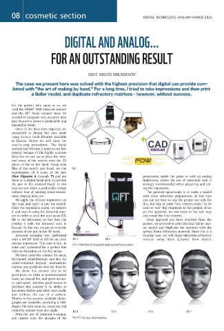
FIG 1-3: Workflow Full digital/Intraoral scanner/Facial scanner
color and optimized for a perfect bite
with no distortion on the full arches.
We have used this scanner for many
full-mouth rehabilitations and also for
screw-retained implant restorations
without any problems with the final fit.
We chose this scanner due to its
good price, no extra or nontransparent
costs, no annual fee, and great accura-
cy and speed. Another good reason to
purchase this scanner is its ability to
document before and after cases easily
and without the use of a camera.
Thanks to this scanner, multiple photo-
graphs are available, providing a 360-
degree file with zoom in, zoom out and
rotatable options from any angle.
FIG 4 FIG 5 FIG 6 FIG 7
With the use of intraoral scanning,
you cannot scan the margins of the FIG 4-7: First step. Facial scanning