


Page 51 - Dental Practice South Asia 18-6
P. 51
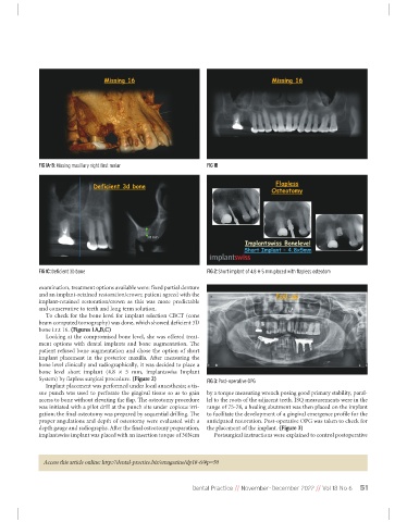
FIG 1A-B: Missing maxillary right first molar         FIG 1B


             FIG 1C: Deficient 3D bone                             FIG 2: Short implant of 4.8 × 5 mm placed with flapless osteotom

             examination, treatment options available were: fixed partial denture
             and an implant-retained restoration/crown; patient agreed with the
             implant-retained  restoration/crown  as  this  was  more  predictable
             and conservative to teeth and long term solution.
                To check for the bone level for implant selection CBCT (cone
             beam computed tomography) was done, which showed deficient 3D
             bone i.r.t 16. (Figures 1A,B,C)
                Looking at the compromised bone level, she was offered treat-
             ment  options  with  dental  implants  and  bone  augmentation.  The
             patient refused bone augmentation and chose the option of short
             implant  placement  in  the  posterior  maxilla.  After  measuring  the
             bone level clinically and radiographically, it was decided to place a
             bone  level  short  implant  (4.8  ×  5  mm,  implantswiss  Implant
             System) by flapless surgical procedure. (Figure 2)    FIG 3: Post-operative OPG
                Implant placement was performed under local anaesthesia; a tis-
             sue  punch  was  used  to  perforate  the  gingival  tissue  so  as  to  gain  by a torque measuring wrench posing good primary stability, paral-
             access to bone without elevating the flap. The osteotomy procedure  lel to the roots of the adjacent teeth. ISQ measurements were in the
             was initiated with a pilot drill at the punch site under copious irri-  range of 75-78, a healing abutment was then placed on the implant
             gation; the final osteotomy was prepared by sequential drilling. The  to facilitate the development of a gingival emergence profile for the
             proper angulations and depth of osteotomy were evaluated with a  anticipated restoration. Post-operative OPG was taken to check for
             depth gauge and radiographs. After the final osteotomy preparation,  the placement of the implant. (Figure 3)
             implantswiss implant was placed with an insertion torque of 30Ncm  Postsurgical instructions were explained to control postoperative


               Access this article online: http://dental-practice.biz/emagazine/dp18-6/#p=50


                                                               Dental Practice // November-December 2022 // Vol 18 No 6  51

