


Page 52 - Dental Practice South Asia 18-6
P. 52
implantology section


                                                                      Manufacturing was done with Sirona CEREC Zirconia A3 meso
                                                                   L block which included milling, sintering and glazing done chairside
                                                                   only on Primemill & Speedfire (Figures 6A,b)
                                                                      Crown  was  cemented  extraorally  over  ti-base  with  dual-cure
                                                                   resin cement.
                                                                      Finally, within 1 hour chair side screw retained prosthesis was
                                                                   prepared which was ready to be delivered. After final placement of
                                                                   prosthesison its desired site, intraorally post-operative radiograph
                                                                   was taken. (Figures 7A,b)
                                                                      Patient was asked to come for timely follow-ups for 3 years, and
                                                                   radiographs were taken respectively, which showed long term suc-
                                                                   cess with short implants. (Figure 8)
                                                                      After  the  3-years  of  follow-up,  CbCT  evaluation  was  done  to
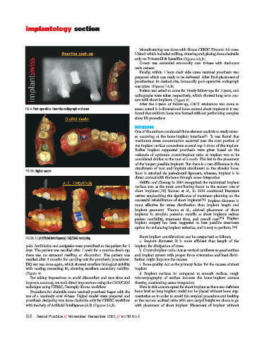
             FIG 4: Post-operative 4months radiograph and scan     assess actual 3-3-dimensional bone around short implant & it was
                                                                   found that uniform bone was formed without performing complex
                                                                   sinus lift procedure.

                                                                   DISCUSSION
                                                                   One of the authors conducted finite element analysis to study stress-
                                                                   es  occurring  at  the  bone-implant  interface [9] .  It  was  found  that
                                                                   maximum stress concentration occurred near the crest portion of
                                                                   the implant surface somewhere around top 5-6mm of the implant.
                                                                   Earlier  implant  supported  prosthesis  were  given  based  on  the
                                                                   rationale  of  optimum  crown/implant  ratio  as  implant  was  to  be
                                                                   considered similar to the root of a tooth. This led to the placement
                                                                   of the longest possible implants. but there is a vast difference in the
                                                                   attachment of root and implant attachment to the alveolar bone.
              FIG 5A: Digital model
                                                                   Root  is  attached  via  periodontal  ligament,  whereas;  implant  is  in
                                                                   direct contact with the bone through osseo-integration.
                                                                      Griffin and Cheung in 2004 recognized the maximized implant
                                                                   surface area as the most contributing factor to the success rate of
                                                                   short  implants.[10]  Romeo  et  al.,  in  2010  conducted  literature
                                                                   review emphasizing the significance of treatment planning on the
                                                                   successful rehabilitation of short implants [11] . Implant diameter is
                                                                   more  effective  for  stress  distribution  than  implant  length  and
                                                                   implant  geometry.  Thoma  et  al.,  advised  placement  of  short
                                                                   implants  in  atrophic  posterior  maxilla  as  short  implants  reduce
                                                                   patient  morbidity,  treatment  time,  and  overall  cost [12] .  Flapless
                                                                   implant  surgery  has  been  suggested  as  best  possible  treatment
                                                                   option for enhancing implant esthetics, and is easy to perform. [13]
                                                                      Short implant considerations can be categorized as follows:
              FIG 5B: A.I (Artificial intelligence) CAD/CAM designing
                                                                      a.  Implant  diameter:  It  is  more  efficient  than  length  of  the
              pain. Antibiotics and analgesics were prescribed to the patient for 5  implant for dissipation of stress.
              days. The patient was recalled after 1 week for a routine check-up;  b. Crown/implant ratio: Act as vertical cantilever so good surface
              there  was  no  extraoral  swelling  or  discomfort.  The  patient  was  and implant system with proper force orientation and load distri-
              recalled after 4 months for carrying out the prosthetic procedures.  bution might improve the success.
              ISQ test was done again, which showed excellent biological stability  c. bone quality: Act as the primary factor for the success of short
              with reading measuring 84, showing excellent secondary stability.  implant.
              (Figure 4)                                              d.  Implant  surface:  As  compared  to  smooth  surface,  rough
                For taking impressions to avoid discomfort and save time and  microtopography  of  surface  increase  the  bone-implant  contact
              improve accuracy, we took direct impressions using the CAD/CAM  thereby, accelerating osseo-integration.
              technique using CEREC, Dentsply-Sirona workflow.        Here in this case we opted for short implant as there was deficient
                Procedure for chairside screw retained prosthesis began with the  bone level so long implant could not be placed without bone aug-
              use  of  a  scanbody  over  ti-base.  Digital  model  were  prepared  and  mentation so in order to avoid this surgical procedure and looking
              prosthesis designing was done chairside only by CEREC workflow  at the narrow occlusal table with zero cuspal height we chose to go
              with the help of Artificial Intelligence (A.I) (Figures 5A,b)  with  placement  of  short  implant.  Placement  of  implant  without

              52   Dental Practice // November-December 2022 // Vol 18 No 6

