Page 13 - DP Vol 20 No 4 HR
P. 13

now an adult—returned with his mother for a review. The long-
           term assessment revealed that the re-attached coronal fragment had
           remained stable and functional over the 8-year period. This case
           underscores that, with appropriate management, the re-attachment
           of a traumatized tooth fragment can result in long-term success and
           durability.


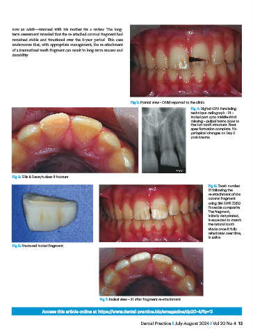
                                                                   Fig 2: Frontal view - Child reported to the clinic.
                                                                                               Fig 4: Digital IOPA Paralleling
                                                                                               technique radiograph - 21 –
                                                                                               Incisal part upto middle-third
                                                                                               missing – pulpal horns close to
                                                                                               the lost tooth structure. Root
                                                                                               apex formation complete. No
                                                                                               periapical changes on Day 6
                                                                                               post trauma.


           Fig 3: Ellis & Davey’s class II fracture

                                                                                                       Fig 6: Tooth number
                                                                                                       21 following the
                                                                                                       re-attachment of the
                                                                                                       coronal fragment
                                                                                                       using 3M ESPE Z350
                                                                                                       Flowable composite.
                                                                                                       The fragment,
                                                                                                       initially dehydrated,
                                                                                                       is expected to match
                                                                                                       the natural tooth
                                                                                                       shade once it fully
                                                                                                       rehydrates over time,
                                                                                                       in saliva.
           Fig 5: Fractured incisal fragment.


                                                    Fig 7: Incisal view – 21 after fragment re-attachment.

                         Access this article online at https://www.dental-practice.biz/emagazine/dp20-4/#p=12

                                                                       Dental Practice I July-August 2024 I Vol 20 No 4  13
   8   9   10   11   12   13   14   15   16   17   18