
Page 14 - DP Vol 20 No 4 HR
P. 14
PEDIATRIC DENTISTRY SECTION
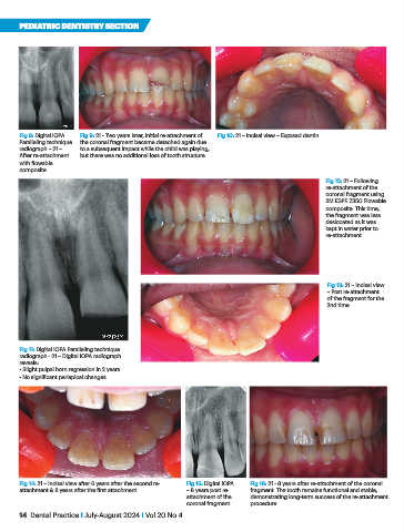
Fig 8: Digital IOPA Fig 9: 21 - Two years later, initial re-attachment of Fig 10: 21 – Incisal view – Exposed dentin.
Paralleling technique the coronal fragment became detached again due
radiograph – 21 – to a subsequent impact while the child was playing,
After re-attachment but there was no additional loss of tooth structure.
with flowable
composite.
Fig 12: 21 – Following
re-attachment of the
coronal fragment using
3M ESPE Z350 Flowable
composite. This time,
the fragment was less
desiccated as it was
kept in water prior to
re-attachment.
Fig 13: 21 – Incisal view
– Post re-attachment
of the fragment for the
2nd time
Fig 11: Digital IOPA Paralleling technique
radiograph - 21 – Digital IOPA radiograph
reveals:
• Slight pulpal horn regression in 2 years.
• No significant periapical changes.
Fig 14: 21 – Incisal view after 6 years after the second re- Fig 15: Digital IOPA Fig 16: 21 - 8 years after re-attachment of the coronal
attachment & 8 years after the first attachment – 8 years post re- fragment. The tooth remains functional and stable,
attachment of the demonstrating long-term success of the re-attachment
coronal fragment. procedure.
14 Dental Practice I July-August 2024 I Vol 20 No 4