
Page 15 - DP Vol 20 No 4 HR
P. 15
II. BIOMIMETIC RECONSTRUCTION POST TRAUMA cluster composites. This approach aimed to achieve a biomimetic
USING MULTI-SHADE NANO CLUSTER COMPOSITES reconstruction that closely mimics natural tooth structure and
A 12-year-old female patient presented to the dental clinic with a aesthetics. Surface characterization, including the replication of
fractured upper left central incisor (tooth number 21). The patient’s enamel lustre and lamellae, was also incorporated into the restoration
father reported that the tooth had sustained a fracture three years ago, process. There are limited articles available regarding restoration of
at age 9, following a fall. The initial injury involved a fracture of the the fractured/ post trauma teeth reconstructions with composites and
distoincisal angle, classified as an Ellis & Davey’s class II fracture. even lesser with nano-cluster composites.
Pre-treatment radiographs indicated adequate dentin thickness.
The tooth was restored using a putty matrix technique, followed by A detailed step-by-step methodology of the reconstruction procedure
incremental layering with enamel, body, and dentin shades of nano- is provided in the following images.
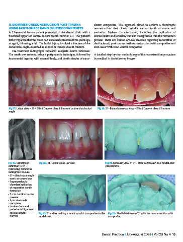
Fig 17: Labial view – 21 – Ellis & Davey’s class II fracture on the distoincisal Fig 18: 21 - Palatal close-up view – Ellis & Davey’s class II fracture.
angle.
Fig 19: Digital high Fig 20: 21– Labial close-up view. Fig 21: Close up view of 21 – after impression and model cast
definition IOPA - preparation.
Paralleling technique
radiograph reveals:
• 21 – distoincisal angle
tooth structure loss
• Regressed pulp
chamber indicative
of reparative dentin
formation.
• 2 mm dentinal barrier
present.
• Apex closure is
complete.
• Lamina dura and
periodontal ligament
spaces appear Fig 22: 21 – after making a mock up with composite on the Fig 23: 21 – Palatal view of 21 with the reconstruction with
normal. model cast. composite.
Dental Practice I July-August 2024 I Vol 20 No 4 15