
Page 13 - DP Vol 22 No 1
P. 13
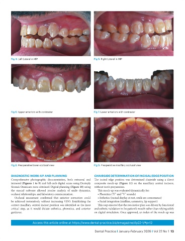
Fig 4: Left Lateral in MIP                             Fig 5: Right Lateral in MIP


           Fig 6: Upper anteriors with contraster                 Fig 7: Lower anteriors with contraster


           Fig 8: Preoperative lower occlusal view                Fig 9: Preoperative maxillary occlusal view


           DIAGNOSTIC WORK-UP AND PLANNING                        CHAIRSIDE DETERMINATION OF INCISAL EDGE POSITION
           Comprehensive photographic documentation, both extraoral and   The  incisal  edge  position  was  determined  chairside  using  a  direct
           intraoral (Figures 1 to 9) and full-arch digital scans using Dentsply   composite mock-up  (Figure 11) on the maxillary central incisors,
           Sirona’s Omnicam were obtained. Digital planning (Figure 10) using   without tooth preparation.
           the exocad software allowed precise analysis of smile dynamics,   This mock-up was evaluated dynamically for:
           occlusal relationships, and laboratory communication.     • Phonetics (“F” and “V” sounds)
              Occlusal assessment confirmed that anterior correction could   • Esthetics (incisal display at rest, smile arc consonance)
           be achieved restoratively without increasing VDO. Establishing the   • Facial integration (midline, symmetry, lip support)
           correct maxillary central incisor position was identified as the most   This step ensured that the restorative plan was driven by functional
           critical step, as it would dictate esthetics, phonetics, and anterior   and esthetic validation in the patient’s mouth rather than relying solely
           guidance.                                              on digital simulation. Once approved, an index of the mock-up was


                          Access this article online at https://www.dental-practice.biz/emagazine/dp22-1/#p=12

                                                                   Dental Practice I January-February 2026 I Vol 22 No 1  13