
Page 15 - DP Vol 22 No 1
P. 15
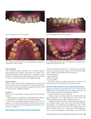
Fig 13: Maxillary anterior tooth preparation          Fig 14: Mandibular anterior tooth preparation


            Fig 15: Maxillary occlusal view of tooth preparation showing vonlays in   Fig 16: Mandibular occlusal view of tooth preparation showing vonlays
            premolars and onlays in molars                        in premolars and onlays in molars


           Anterior segment:                                      Chairside provisional restorations were fabricated using 3M Espe
           Ultra-conservative veneer preparations were performed using the   Protemp 4, a bisacryl temporary using the index of the printed digital
           mock-up-guided APT technique. Preparations were confined to the   mock-up design. This phase allowed the assessment of:
           enamel. Incisal edges were left unprepared, as the length was added   • Functional comfort
           restoratively. Minimal mesial margin opening was performed on teeth   • Speech adaptation
           11 and 12 to correct the midline deviation. (Figures 12 to 14)  • Esthetic acceptance
                                                                  • Occlusal stability
           Posterior segment:                                        The patient reported immediate improvement in function and
           Adhesive onlays were planned to integrate posterior occlusion with   appearance, with no muscular discomfort or phonetic issues.
           the new anterior scheme. Second molars were excluded and served as
           stable posterior stops, allowing efficient preparation without the loss   DEFINITIVE RESTORATIONS AND ADHESIVE PROTOCOL
           of occlusal reference. (Figures 15 and 16)             Definitive restorations were fabricated in IPS e.max, Lithium disilicate
                                                                  (Figure 18) due to its favourable esthetic and adhesive properties. Lab
           Exception:                                             work was done by Precision Dental Studio Ltd (Mumbai).
           Teeth 32 and 33 required full-coverage restorations due to functional   A standard adhesive protocol was followed:
           demands.                                                  • Veneer: Veneer was etched using Ivoclar Monobond Etch and
              All preparations were completed in a single appointment, supported   Prime (60 seconds). It is a single component ceramic primer that
           by a stable posterior occlusion and mock-up-guided planning.   etches and silanates the glass-ceramic surfaces in one step.
              The soft tissue was managed using the retraction paste by 3M. The   • Teeth: Teeth isolation was done with a split dam and temporaries
           preparation was scanned using Dentsply Sirona’s Omnicam scanner.   were carefully removed. Teeth were etched using 38% phosphoric
           And it was sent to the lab where final designing was done based on the   acid (Pulp dent, USA) followed by Bond application (Tetric N bond
           provisionals. (Figure 17)                              universal, Ivoclar Vivadent). Restorations were bonded using a resin
                                                                  cement (Variolink Esthetic LC, neutral shade). After bonding, the
           PROVISIONALIZATION AND PATIENT ADAPTATION              excess cement was immediately removed using a micro brush before

                                                                   Dental Practice I January-February 2026 I Vol 22 No 1  15