
Page 15 - Dental Technology June 2022
P. 15
14-17-Nico article-Q8_6-7-8-Ivoclar.qxd 7/13/2022 9:10 PM Page 2
laboratory section 15
DENTAL TECHNOLOGY, APRIL-JUNE 2022
Air nd 1,00
Enamel nd 1,63
DEC nd 1,43
Dentin nd 1,54
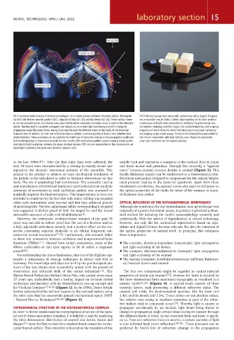
FIG 4: A natural tooth consists of three essential layers, all of which possess different refractive indices: The enamel FIG 5: Waxing-up exercises using tooth-colored wax set by August Bruguera
(nD 1.64), the dentino-enamel junction (DEJ), depicted in blue (nD 1.43) and the dentin (nD 1.54). These indices, howev- are an excellent way to foster a better understanding of the histo-anatomi-
er, are not absolute values, but relative ones given that localized variations commonly occur in each of the refractive cal influence on tooth color and enable its imitation. The professional wax
indices. Thereby, light is caused to propagate non-linearly by an incident light beam being forced to change its set includes modeling and effect waxes, the excellent modeling and scraping
propagation angle (θ) several times during its journey through the different layers of the tooth. At the transition properties of which allow the dental technician quick and clean contouring
from one layer to another, i.e. from one refractive index to another, a certain quantity of light is also reflected back and shaping of any model waxup. Thanks to the balanced flow and solidifica-
(total reflection). These processes can be predicted by Snell's law of refraction (change in the propagation angle) and tion time in conjunction with ideal stability, even filigree structures like
by Fresnel equations. In the natural enamel, the very small (< 260 nm) hydroxyapatite crystals create a bluish, opales- cusps and mamelons can be shaped precisely.
cent (elastic) light scattering, whereas the larger dentinal tubules (>780 nm) are responsible for the characteristic yel-
lowish light scattering that gives each tooth its intrinsic color.
to the late 1960s [4,5] . After the first static data were collected, the middle third and represents a transition to the occlusal third in upper
next 20 years were characterized by a striving to exactly record and and lower molars and premolars. Through this concavity, a “sigmoid
reproduce the dynamic movement patterns of the mandible. This curve” (convex enamel/concave dentin) is created (Figure 3). This
peaked in the attempt to achieve an exact mechanical simulation of locally thickened enamel can be understood as a biomechanical rein-
the patient in the articulator in order to fabricate restorations on that forcement mechanism designed to compensate for the typical, height-
basis. The era of gnathology had commenced. The successful tracing ened pressure loading in the posterior quadrants. Apart from these
and reproduction of individual functional and nonfunctional condylar mechanical correlations, the sigmoid curves also exert an influence on
pathways of movement in each individual patient was supposed to the optical properties of the tooth: by virtue of the variance in enam-
markedly improve the final restoration. This improvement in turn was el thickness they define.
intended to stand out by the fact that only minor milling was required
when such restorations were inserted and that they achieved greater OPTICAL INFLUENCES OF THE HISTOANATOMICAL MORPHOLOGY
clinical longevity. The less adjustment effort correspondingly required Although the motivation for the biomechanical wax-up technique was
was supposed to deliver proof of both the hoped-for and the actual originally of a purely functional nature, in practice it provided a didac-
successful outcomes of such oral rehabilitations [6] . tical method for imitating the tooth’s exomorphology naturally and
However, the systematic, evidence-based research of the past 15 aesthetically. With the advent of digitalization in dental technology,
years was not able to deliver proof that the use of a facebow and/or however, not only did the anatomical reproduction based on algo-
a fully adjustable articulator actually had a positive effect on the cor- rithms and digital libraries become relevant, but also the imitation of
rective contouring required chairside or on clinical long-term out- the optical properties of natural teeth. In principal, this comprises
comes for dental restorations [7-10] . Furthermore, this research failed three components:
to show any connection between occlusion and temporomandibular
disorders (TMDs) [11] . Viewed from today's perspective, some of the ♦ The complex, direction-dependent (anisotropic) light propagation
efforts undertaken at that time appear to be of rather a dogmatic and light scattering of the dentin
nature [12] . ♦ The complex, direction-independent (isotropic) light propagation
Notwithstanding the above limitations, that era of the Eighties rep- and light scattering of the enamel
resents a renaissance in wax-up techniques in dental tech labs in ♦ The varying volumetric distribution behaviours (different thickness-
Germany. The knowledge and ideas set forth by the gnathological pio- es) between dentin and enamel
neers of the late Sixties were successfully united with the powers of
observation and technical skills of the dental technician [13] . The The first two components might be regarded as optical material
Master Dental Technician Michael Heinz Polz, who passed away some properties of dentin and enamel [22] , whereas the latter is dictated by
17 years ago, undoubtedly had a lasting impact on German dental the three-dimensional histo-anatomical topography as visualized in a
technology and dentistry with his biomechanical wax-up concept and natural tooth [21,23] (Figure 4). A natural tooth consists of three
his "Occlusal Compass” [14-15] (Figure 1). In the 1990s, Dieter Schulz essential layers, each possessing a different refractive index: The
further systematized the defined regions on the occlusal surface using enamel (nD 1.64), the dentinoenamel junction, DEJ for short (nD
the color code that has meanwhile gained international repute (NWT 1.43) and the dentin (nD 1.54). These indices are not absolute values,
– Natural Wax-up Technique) [16-20] (Figure 2). but relative ones seeing as localized variations in each of the refrac-
tive indices tend to commonly occur [24] . Thereby, light is caused to
TOPOGRAPHICAL STRUCTURE OF THE HISTOANATOMICAL COMPLEX propagate non-linearly by an incident light beam being forced to
In order to better understand the topographical structure of the natu- change its propagation angle several times during its journey through
ral tooth’s histo-anatomical complex, it is helpful to start by analysing the different layers of tooth. At the transition from one layer to anoth-
the three-dimensional distribution of enamel and dentin. Bazos and er, i.e. from one refractive index to another, a certain quantity of light
Magne [21] were the first to describe a marked dentin concavity on the- is also reflected back (total reflection) [24-26] . These processes can be
upper buccal surface. This concavity is located at the transition of the predicted by Snell's law of refraction (change in the propagation