
Page 16 - Dental Technology June 2022
P. 16
14-17-Nico article-Q8_6-7-8-Ivoclar.qxd 7/13/2022 9:10 PM Page 3
16 laboratory section DENTAL TECHNOLOGY, APRIL-JUNE 2022
angle) and by Fresnel equations. In the nat- exerts a significant influence on the lumi-
ural enamel, the very small (< 260 nm) nous reflectance (brightness) of natural THE HISTO-ANATOMICAL WAX-UP TECHNIQUE
hydroxyapatite crystals create a bluish, tooth. Early studies by Kraus [30] (1952) Waxing-up exercises using tooth-colored wax
opalescent (elastic) light scattering whereas showed that the enamel at the bucco- are an excellent way to foster a better under-
the larger dentinal tubules (> 780 nm) [27] occlusal surfaces of the first lower molars standing of the histo-anatomical influence
are responsible for the characteristic yellow- demonstrates the largest enamel thickness of on tooth color while enabling its imitation as
ish light scattering each of which gives the all dentition in the jaw (1.8 to 2.2 mm). That well. The professional wax set by August
tooth its intrinsic color [28] . is why – absent all abrasion or erosion phe- Bruguera allows quick and clean contouring
In this connection, it is interesting to note nomena – the lower first molar usually of any wax-up using modeling and effect
that the literature often reports that tooth shows the highest luminance of all teeth. waxes that exhibit excellent modeling and
color is predominantly dictated by the dentin. The ratio of coronal configuration to enamel scraping properties. Thanks to the balanced
This notion appears to confirm the common thickness (three components) is particularly flow and solidification time partnered with
perception that the translucence from natural reflected in the most recent approaches an ideal stability, even filigree structures like
enamel is higher than from natural dentin. found in digital reproduction [31] . In this con- cusps and mamelons can be shaped precise-
Yet such a statement is not entirely true. A text, the striving is to reproduce such histo- ly (Figure 5). The goal is not only to imitate
study by Yu et al [29] showed that the average anatomical restorations completely by the exomorphology of the posterior teeth
translucence of enamel is only slightly higher mechanical means. A hybridization of sub- based onthe natural model – as is the tradi-
(approx. 2%) than that of dentin. tractive (CAD/CAM-supported milling) and tional convention – but also to reproduce the
The fact that translucence is a function of additive (3D printing) fabrication technolo- internal endomorphology of the dentin in
a tooth’s thickness and that dentin is usually gy is designed to achieve this [32] . At present, order to enable a better understanding of the
thicker than enamel has frequently been to what extent this will actually be feasible in optical interaction of both components
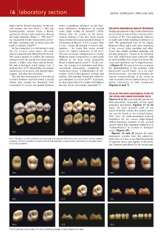
overlooked. As a result, the enamel thickness the near future still remains uncertain [33] . (Figures 6 and 7).
ATLAS OF THE HISTO-ANATOMICAL STUDY OF
THE UPPER AND LOWER POSTERIOR TEETH
Figures 8 to 16 systematically present the
histo-anatomical topography of the upper
premolars and molars. Figures 17 to 25
depict the lower posterior teeth. In our
model of Mother Nature, the sigmoid curves
can also be found on the occlusal surface.
They form the histo-anatomical dentinal
foundation for the convex, bulge-shaped,
shifted functional elements of the occlusal
surface which Polz sometimes referred to as
"Rucksackhöcker” in German or “backpack
cusps” (Figure 25).
FIG 6 FIG 7
Figures 16 and 25 present the histo-
anatomical complex from the palatal/lin-
FIG 6-7: The goal is not only to imitate the exomorphology of the posterior teeth based on the natural model – as is the traditional gual. Here, it is easy to see that the principle
convention – but also to reproduce the dentin’s internal endomorphology to enable a better understanding of the optical interac- of sigmoid curves also applies in the proxi-
tion of both components.
mal direction and allows for the strong mar-
FIG 8 FIG 9 FIG 10
FIG 11 FIG 12 FIG 13
FIG 14 FIG 15 FIG 16
FIG 8-16: Systematic representation of the histo-anatomical topography of upper premolars and molars.