
Page 10 - DT14-1
P. 10
10-12-Domenico Benagiano_6-7-8-Ivoclar.qxd 4/15/2024 7:57 AM Page 1
10 implantology section DENTAL TECHNOLOGY, JANUARY-MARCH 2024
A COMPREHENSIVE ANALYSIS OF DIGITAL VS.
CONVENTIONAL TECHNIQUES IN DENTAL IMPLANTOLOGY
DOMENICO BENAGIANO, DDS AND CLAUDIA SALERNO, DDS
In this article we show a prosthesis implant rehabilitation clinical case of the lower arch through a fully digital
CAD-CAM workflow. We opted to carry out a cobalt-chromium-ceramic rehabilitation due to the structural and aesthetic
benefits of this combination confirmed by decades of clinical case studies. The technologies available for dental units,
dental laboratories involved and production centres allowed to apply the fully digital workflow at each step.
INTRODUCTION
The analogue or conventional prosthesisation on implants provides
for the steps of registering an alginate impression, creating an indi-
vidual impression tray, second impression registration session with
implant transfer using the individual impression tray and silicone
material and face-bow registration, development of the cast or resin
model with similar implants, fitting in articulator, diagnostic wax-
ing, position test in the oral cavity, conversion of the diagnostic
waxing in small structure, production of this structure through lost-
wax casting technique, structure test in the oral cavity, biscuit test,
then finalisation and delivery. Compared to this workflow, the fully
digital technique offers a path consisting of fewer steps: detection
of the optical impression with scan abutments in the oral cavity,
CAD modelling of the final anatomy and CAM production of the
anatomy and of the structure made of temporary material, test of
the aluminium structure so as to verify the fit on the implants and
depending on the aesthetic test of the anatomy which is then deliv-
ered based on a temporary function which can be functionalised,
registering the functionalised anatomy by 3D scanning.
This information is then sent to the laboratory, the final prosthe-
sis is made using the CAD-CAM technology in compliance with the
functional occlusal plane, delivery of the finished and already func-
tionalised work.
Given that each test and each assessment carried out by the
physician is performed with test specimen made of cost effective
material, fully compliant in the shape and in the overall dimensions
of the prosthesis being created in the dental laboratory, the labora-
tory makes the final prosthesis without interruptions and without
dispatch, as well as products recovery times.
This allows the patient to retain and use the test specimen tem-
porarily, without hindering or slowing down the work of the labo-
ratory. Each change or correction is digitally communicated by the
dentist’s office to the laboratory and vice versa allowing a coordi-
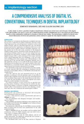
nated and, above all, continuous and uninterrupted workflow. FIG 1
MATERIALS AND METHODS
Prosthesisation was performed on Branemark Ø4.1-compatible
external hexagonal connection implants.
A Trios 3 Pod intraoral scanner manufactured and sold by
3Shape was used.
The scan abutments were designed, developed, and manufac-
tured by TQM srl.
CAD modelling and aesthetic finalisation were carried out by the
dental laboratory Zirkonlab while exocad version Galway software
was used for CAD modelling.
The aluminium verification jig, the full PMMA anatomy and the
final chromium-cobalt structure were manufactured by the TQM
production facility.
Mikron HSM200U LP was the machine used for milling metal
components, while the PMMA anatomy copy was made using a
model DWX-51D Roland milling machine.
HyperDENT version 8.2 was the CAM software used for all
milling productions. The aluminium used for the trial structure is
Magnum Ergal manufactured by MESA.
The Chromium-Cobalt used for the final structure is Magnum FIG 2