
Page 15 - DT14-1
P. 15
14-20-Mohit-Q8_6-7-8-Ivoclar.qxd 4/15/2024 8:01 PM Page 2
DENTAL TECHNOLOGY, JANUARY-MARCH 2024
prosthetic section 15
MART) (Figures 10 to 14). The mock-up allowed for patient feedback
and consent regarding the anticipated changes in tooth morphology, func-
tional occlusion, and aesthetics. Following patient counselling and agree-
ment, the treatment proceeded with a focus on minimally invasive
occlusal overlays and veneers wherever possible.
PREPARATION
Initially, at the first visit for teeth preparation, both the upper and lower
teeth from 1st premolar to 1st premolar were temporized over non-pre-
pared teeth. This was done so as to create the designed vertical stop of the
newly created occlusal scheme.
Later, the upper and lower posterior teeth from 2nd premolar to 2nd
molar were prepared with the decided design for veneerlays and overlays
(Figures 15 to 18). These designs were specifically chosen for a mini-
mally invasive, non-retentive occlusal coverage where all the margins
were supragingival.
The supragingival margin was chosen for two reasons:
1. To facilitate easier isolation and bonding by preventing crevicular
fluid seepage and inflammation during the temporization phase.
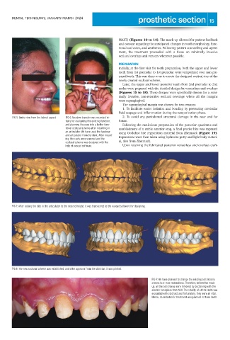
FIG 5: Smile view from the lateral aspect FIG 6: Facebow transfer was recorded ini- 2. To avoid any periodontal structural damage in the near and far
tially for evaluating the existing function, future.
and planning the case into a better func- Following the meticulous preparation of the posterior quadrants and
tional occlusal scheme after mounting in establishment of a stable anterior stop, a final precise bite was captured
an articulator. We have used the facebow using Occlufast bite registration material from Zhermack (Figure 19).
and articulator from Corident. After mount-
ing, the casts were scanned and the Impressions were then taken using hydrorise putty and light body materi-
occlusal scheme was designed with the al, also from Zhermack.
help of exocad software. Upon receiving the fabricated posterior veneerlays and overlays craft-
FIG 7: After raising the bite in the articulator to the desired height, it was transferred to the exocad software for designing.
FIG 8: The new occlusal scheme was established, and after approval from the clinician, it was printed.
FIG 9: We have planned to change the existing old zirconia
crowns to e-max restorations. Therefore, before the mock-
up, all the old crowns were removed by sectioning with the
electric handpiece from NSK. The vitality of all the teeth was
evaluated with cold test and fortunately, they were all vital.
Hence, no endodontic treatment was planned in those teeth.