
Page 32 - DT Vol 15 No 3
P. 32
32 I implantology DENTAL TECHNOLOGY, JULY-SEPTEMBER 2025
DIGITALLY GUIDED IMMEDIATE FULL-ARCH
REHABILITATION WITH CUSTOMIZED 3D-PRINTED
SHELL TEMPLATES: A CASE REPORT
ANAND KRISHNAMURTHY, RAHUL KAKODKAR, RONIL KAKODKAR
INTRODUCTION
Full-arch implant rehabilitation with immediate loading offers functional,
psychological, and esthetic benefits to edentulous patients or to those with
terminal dentition. Predictability of such treatments relies on accurate implant
positioning, maintenance of vertical dimension, and a passive-fitting definitive
prosthesis. The integration of digital workflows and additive manufacturing has
enabled clinicians to streamline this process, enhancing precision and reducing
the treatment time. This report details a workflow incorporating a 3D-printed shell
used as a customized special tray to capture implant positions and verify occlusion
simultaneously, ensuring a verified master cast for passive bar fabrication and
timely delivery of the final zirconia restoration.
CASE PRESENTATION
A 52-year-old female presented with advanced periodontitis, severe bone loss,
and generalized mobility with esthetic and functional impairment. The patient
requested fixed rehabilitation with immediate function.
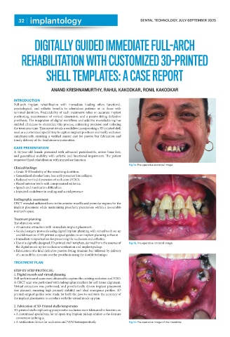
Fig 1a: Pre-operative extraoral image
Clinical findings:
• Grade II-III mobility of the remaining dentition.
• Generalized alveolar bone loss with posterior bite collapse.
• Reduced vertical dimension of occlusion (VDO).
• Flared anterior teeth with compromised esthetics.
• Speech and mastication difficulties.
• Impaired confidence in smiling and social presence
Radiographic assessment:
CBCT revealed sufficient bone in the anterior maxilla and premolar regions for the
implant placement while maintaining prosthetic parameters within a favourable
interarch space.
Treatment planning:
The objectives were:
• Atraumatic extraction with immediate implant placement.
• Guided surgery protocols using digital implant planning with virtual teeth set up
and fabrication of 3D printed surgical guides on an implant planning software
• Immediate temporization for preserving the occlusion and esthetics.
• Use of a digitally designed 3D-printed shell template, derived from the essence of Fig 1b: Pre-operative intraoral image
the digital mock-up for occlusion verification and implant pickup.
• Fabrication of a final definitive passive-fitting titanium bar followed by delivery
of a monolithic zirconia overlay prosthesis using the double technique
TREATMENT PLAN
STEP-BY-STEP PROTOCOL:
1. Digital records and virtual planning
Full-arch intraoral scans were obtained to capture the existing occlusion and VDO.
A CBCT scan was performed with radiographic markers for soft tissue alignment.
Virtual extraction was performed, and prosthetically driven implant placement
was planned, ensuring high primary stability and ideal emergence profiles. 3D
printed surgical guides were made for both the jaws to maintain the accuracy of
the implant placements to correlate with the virtual mock up plan.
2. Fabrication of 3D-Printed shells/temporaries
3D-printed shells replicating preoperative occlusion were fabricated to function as:
• A customized special tray for an open-tray implant pickup similar to the denture
conversion technique.
• A verification device for occlusion and VDO intraoperatively. Fig 1c: Pre-operative image of the mandible