
Page 32 - DP20-2.qxd
P. 32
MAGNIFICATION seCTION
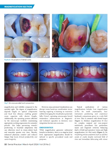
Fig 6a-b: Visualization of dental caries
Fig 7: Microscope aided tooth preparation
magnification and visibility compared to the However, unaccustomed visualizations can Typical applications of various
unaided sight. The degree of magnification disrupt innate hand-eye coordination motor magnifications include: Low magnification
depends largely on optical components learning requiring re-training to maximize (3–8x) for tooth examination, bur/
and focal lens distance enabling greater utilities leveraging the visualization potentials instrument positioning and anatomical
zoom capacities with shorter lengths. fully. Overall, operating microscopes herald landmark comparisons given in a wide field
Additionally, the operating posture afforded tremendous enhancements in diagnostic of view. This is common with dental loupes
by the microscope facilitates maintaining and technical capacities in dentistry once (Figure 1); Medium magnification (8–16x)
appropriate working ergonomics for extended integration barriers are overcome. for complex surgical and non-surgical
periods. This helps preventing stressful endodontic procedures like perforation
positioning or hunched adaptations that ENDODONTICS repair and fragment retrieval, balancing
can otherwise result in strain-related back While magnification generates substantial depth of field and operatory vision and High
and muscular spasms over time. Thereby, benefits in dentistry, there is no singular ideal magnification (16–30x) rarely (Figure 2), for
enhanced physician comfort and reduced level. Appropriate magnification should be visualizing minute anatomies such as calcified
fatigue help boost clinical productivity and tailored to specific procedural needs and canals or cracks despite restricted field of
career longevity. purposes. view and immediate defocusing with minor
32 Dental Practice I March-April 2024 I Vol 20 No 2