
Page 33 - DP20-2.qxd
P. 33
with coaxial lighting, it enables detecting
preliminary paths for removable partial
denture placement while minimizing
long-term abutment damage. For fixed
prosthodontics, horizontal and vertical
marginal fits between prostheses and
abutments impact the prognosis, durability
and aesthetics. Operating microscopy reveals
microscopic marginal ridge elevations
and margins above preparation finishes
unapparent to the naked eye. This allows
selective adjustments for incremental
fit improvements when seating indirect
restorations.
Ideally, the horizontal gap between
dental prostheses and abutments is 0 μm,
readily achievable through magnification,
laboratory precision and training. However,
the optimal vertical gap is 50μm, which is
influenced by preparation, impressions and
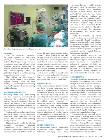
Fig 8: Operating microscopes in Maxillofacial surgeries materials. Minimizing marginal discrepancies
considerably reduces marginal decay and
movements. fissures (Figure 5), inspecting caries borders/ improves patient comfort.
Since its emergence, endodontics remaining decay (Figures 6a and 6b), Oral proprioception senses thickness
has greatly capitalized on magnification verifying matrix adaptation and controlled or roughness differences less than 20μm.
advantages. Conventional benefits liner application, conservative preparation of Obtaining smooth prosthesis surfaces through
include Identifying/accessing anatomical small Class III cavities, evaluating restorative precise placements and comprehensive
complexities, negotiating sclerosed canals, margins, detecting material gaps, voids or polishing is paramount for health and
visualizing dystrophic calcifications for impurities, removing excess composites comfort. Magnification enables enhanced
removal, improved obturation and coronal and managing accidental pulp exposures to precision in prosthodontic evaluations
seals, detecting/repairing defects like prevent mechanical injury. and procedures, allowing detections and
perforations (Figures 3a and 3b), retrieving Microscopes and loupes upgrade direct refinements unattainable by unaided vision.
separated instruments or posts. diagnostic capacity and precision in various
Many clinicians now utilize microscopes facets of tooth-preserving restorative PERiODONTiCS
in periradicular (Figure 4) and periodontal treatments through enhanced visualization. Thorough diagnosis in periodontal treatments
surgeries, enabling delicate soft tissue necessitates meticulousness, best achieved
handling and accurate replacement resulting ORTHODONTiCS through microscopy and micro-instruments,
in reduced surgical morbidity, less scarring, While adoption remains limited presently, enabling minimally invasive techniques.
and faster healing. the dental operating microscope shows The dental operating microscope facilitates
promise in orthodontics warranting further comprehensive identification of periodontal
RESTORATiVE DENTiSTRY investigations. Existing evidence indicates irritants like calculus or enamel projections,
Diagnosing dental caries relies heavily that microscopy significantly enhances the explaining localized attachment loss,
on visual inspection, supplemented by debonding and cleanup outcomes regardless especially in hindered sites like furcations.
radiographs for confirmation. Evidence of the tools and finishing methods chosen, In recent years, various microsurgical
indicates that tactile examination does not by enabling superior visualization of approaches have been developed with
improve the accuracy of caries diagnosis. microscopic details. One critical advantage excellent outcomes and patient satisfaction.
Detecting early non-cavitated lesions enables is conserving enamel during adhesive Documented benefits encompass predictable
remineralization therapies thus preserving removal following bracket detachment. The results, minimally invasive procedures with
the tooth structure. magnification enables differentiation and less discomfort, expedited healing, improved
Studies reveal low to medium magnification selective elimination of bonding material esthetics and high patient acceptance.
significantly enhances diagnosis as compared versus enamel that is indistinguishable by Micro-principles are commonly applied
to the direct vision. Lower levels maintain direct vision. Thereby, loss of enamel structure when incising or suturing periodontal flaps
adequate field of view without sacrificing and associated sequelae can be minimized or by enabling controlled, trauma-free soft
depth perception. prevented. tissue manipulation without stretching,
Magnification also benefits the following distorting or tearing. Microscopy upgrades
restorative procedures; identifying PROSTHODONTiCS direct visualization to transform periodontal
demineralized enamel, visualizing old In prosthodontic practice, medium-level therapy - early disease detection, conservative
restorations for minimally invasive removal, magnification (6-8x) simplifies many interventions with shorter recovery, and long-
detecting enamel and dentinal cracks and technically challenging tasks. Along term restoration of form and function.
Dental Practice i March-April 2024 i Vol 20 No 2 33