Page 43 - DP20-2.qxd
P. 43

complex case report of oral rehabilitation in a
                                                                                     young male after a successful and conservative
                                                                                     management  of  extensive  ameloblastoma  in
                                                                                     the mandible, with the placement of three
                                                                                     Swiss implants and lower prosthesis fabricated
                                                                                     with the help of a digital workflow.

                                                                                     CASE REPORT
                                                                                     21-year-old male reported to the clinic with
                                                                                     the chief complaint of swelling on one side
                                                                                     of the face for the last 3 years. Currently,
                                                                                     he was experiencing pain and was unable
                                                                                     to eat. He had previously consulted plastic
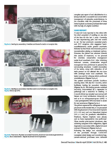
           Fig 2a-b: Healing by secondary intention and bone formation at surgical site  and ENT surgeons, who suggested hemi-
                                                                                     mandibulectomy under general anesthesia
                                                                                     followed by free fibula microvascular graft or
                                                                                     reconstruction plating  as  treatment options.
                                                                                     However, the patient was reluctant towards
                                                                                     this course of treatment, so we planned
                                                                                     for chairside conservative management
                                                                                     under local anesthesia (LA). After obtaining
                                                                                     informed consent, conservative surgical
                                                                                     management was performed to preserve the
                                                                                     surrounding structures. Careful enucleation
                                                                                     of  the  lesion,  along  with  extraction  of  all
                                                                                     teeth present on the affected  site was done
                                                                                     with curettage under local anesthesia. The
                                                                                     lesion was sent for a biopsy, which confirmed
                                                                                     ameloblastoma (Figures 1a, b, c).
                                                                                     The patient underwent periodic iodoform
                                                                                     dressing every 10th day for 3 months, which
                                                                                     facilitated  healing  by secondary  intention
                                                                                     (Figures 2a, b). The healing process unfolded
           Fig 3a-b: Healing by secondary intention and bone formation at surgical site  smoothly, characterized by a regression in
           Fig 3c: Follow-up OPG
                                                                                     pathology and a reduction in swelling. A
                                                                                     6-month follow-up revealed near-normal soft
                                                                                     tissues and bony architecture intraorally as
                                                                                     well as in an orthopantomogram (OPG). A
                                                                                     1-year postoperative OPG was taken to assess
                                                                                     for any recurrence (Figures 3a, b, c).
                                                                                     The patient expressed his willingness for teeth
                                                                                     placement at the treated site, so we proceeded
                                                                                     with the placement of two bone-level implants
                                                                                     (3.3 x 10 mm) and one tissue-level implant
                                                                                     (4.8 x 6 mm) for prosthetic rehabilitation.
                                                                                     Freehand,  flapless  implants were  placed,
                                                                                     and no bone augmentation was performed.
                                                                                     Three  months  post-implant  placement,  after
                                                                                     achieving adequate Osseo integration, digital
                                                                                     impressions were taken with a digital intraoral
                                                                                     scanner (Primescan) (Figures 4a, b, c).
                                                                                     Following  the  design  and  manufacturing
                                                                                     of the prosthesis through CAD/CAM
           Fig 4a-b: Free hand, flapless bone level implants placement (no bone augmentation)  (computer-aided design and manufacturing),
           Fig 4c: Stock abutments – digital abutment level impression               cement-retained prostheses were delivered

                                                                       Dental Practice i March-April 2024 i Vol 20 No 2  43
   38   39   40   41   42   43   44   45   46   47   48