


Page 55 - DP Vol 20 No 3
P. 55
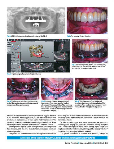
Fig 2: Initial radiographic situation, before loss of 33, 31, 41  Fig 3: Pre-surgical clinical situation.


                                                                                 Fig 5: Positioning of the guides. The primary part,
                                                                                 blue in color, is not removed until the end of the
                                                                                 surgical and prosthetic procedures.

            Fig 4: Digital design of prosthetic implant therapy.


           Fig 6: The implants with the mounters at the   Fig 7: Implants inserted after removal of   Fig 8: The placement of the reinforced
           end of the positioning, guided by the second   the portion of template used for implant   provisional on the prosthetic cylinders. The
           component of the guide, purple.      placement. The accessibility of the operating   position of the occlusal plane is exactly what
                                                field allows the management of soft and hard   was defined in the digital design phase.
                                                tissues with access completely equivalent to
                                                an open field surgery.

           elements in the anterior sector, recently lost the last support elements   to the early loss of dental elements and the use of removable dentures
           of the lower arch. In the upper arch, the patient already had a fixed   for many years. Additionally, the patient had a small thickness of
           prosthesis on implants made 10 years earlier. Following the loss of the   keratinized tissues.
           remaining lower dental elements due to marginal infiltration, it was   In contrast to the upper arch, which was treated few years back
           necessary to convert the lower prosthesis into a complete denture.  with unguided surgery and prosthetic immediate loading using the
              At the patient’s request, a new lower prosthesis was designed on   “One Model” technique, we opted for an entirely digital design and
           fixed implants, with the same characteristics as the upper prosthesis   implementation for the lower arch, utilizing guided surgery with the P
           made few years back.                                   cube method (Oxy Digital Solutions, Biomec).
              The patient had limited bone volumes in the posterior sectors due   The patient underwent an intraoral scan (Trios 3, 3Shape) of

                         Access this article online at https://www.dental-practice.biz/emagazine/dp20-3/#p=52

                                                                         Dental Practice I May-June 2024 I Vol 20 No 3  53

