


Page 59 - DP Vol 20 No 3
P. 59
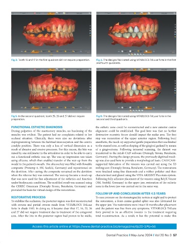
Fig 3: Tooth 16 and 17 in the first quadrant did not require preparation.  Fig 4: The designs fabricated using VITABLOCS TriLuxe forte in the first
                                                                   and fourth quadrants.


           Fig 5: In the second quadrant, tooth 25, 26 and 27 did not require   Fig 6: The designs fabricated using VITABLOCS TriLuxe forte in the
           preparation.                                            second and third quadrants.

           funCTIOnAL eSTheTIC dIAGnOSIS                          the esthetic zone could be reconstructed and a new anterior canine
           During palpation of the masticatory muscles, no hardening of the   alignment could be established. The goal here was that no further
           muscles was evident. The patient had no complaints related to her   destructive eccentric forces should impact the molar area. The first
           occlusal situation. Clinically, there  were also no deviations after   step was restoration of the upper anterior region. Following local
           deprogramming between the habitual intercuspation and the centric   anesthetic, the mock-up supported gentle preparation that was limited
           condyle position. There was only a loss of vertical dimension as a   to the enamel area, as well as shaping of the gingival garland by means
           result of abrasive and erosive processes. For this reason, the bite was   of a gingivectomy. Following intraoral scanning, the dataset was
           raised by one millimeter in the articulator in order to be able to carry   transferred to the inLab CAD software (Dentsply Sirona, Bensheim,
           out a functional esthetic wax-up. The wax-up impression was taken   Germany). During the design process, the previously digitized mock-
           using silicone, which then enabled transfer of the wax-up from the   up was also used here to provide a morphological basis. CAD/CAM-
           model to the patient’s mouth. The silicone key was filled with flowable   supported  fabrication  of  the  veneers  was  carried  out  using  the  X5
           composite (Protemp 4, 3M, Seefeld, Germany) and repositioned on   milling unit (Dentsply Sirona, Bensheim, Germany). The restorations
           the dentition. After curing, the composite remained on the dentition   were finalized using fine diamonds and a rubber polisher and then
           when the silicone key was removed. The wax-up became a mock-up   characterized and glazed using the VITA AKZENT Plus stain system.
           that was now used for fine adjustment of the esthetics and function   Following fully adhesive placement of the veneers using RelyX Veneer
           under biodynamic conditions. The modified result was scanned using   (3M, Seefeld, Germany) in the upper jaw, restoration of the esthetic
           the CEREC Omnicam (Dentsply Sirona, Bensheim, Germany) and   zone in the lower jaw was carried out in the same way.
           provided the basis for virtual design of the restorations.
                                                                  fOLLOw uP And COnCLuSIOn AfTer 4.5 yeArS
           reSTOrATIVe PrOCeSS                                    To ease pressure on the stomatognathic system at night and to protect
           To stabilize the occlusion, the posterior region was first reconstructed   the restoration, a front-canine-guided splint was also fabricated for
           with  crowns  and  partial  crowns  made  from  VITABLOCS  TriLuxe   the upper jaw. The restorations were intact 18 months after placement
           forte in shade 1M2. In doing so, it became clear that 17, 16, 25, 26   and the patient was free from any symptoms. VITABLOCS TriLuxe
           and 27 did not require treatment due to treatment of the antagonist   forte proved to be an effective booster in the treatment requiring
           area. After the bite in the posterior region had proven to be stable,   total reconstruction. As a result, it has the potential to make this


                         Access this article online at https://www.dental-practice.biz/emagazine/dp20-3/#p=56

                                                                         Dental Practice I May-June 2024 I Vol 20 No 3  57

