
Page 26 - DP Vol 20 No 4 HR
P. 26
ORTHODONTIC SECTION
TRANSFORMATIVE TREATMENT:
ORTHO AND ORTHOGNATHIC SURGERY
Shivani Patel, Paul Johnson and Raul Costa
The authors present their transformative treatment case, an award-winning case from
Dentistry Clinical Case Awards 2022
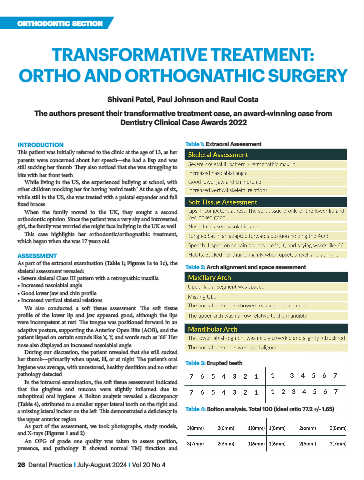
INTRODUCTION Table 1: Extraoral Assessment
This patient was initially referred to the clinic at the age of 13, as her
parents were concerned about her speech—she had a lisp and was
still sucking her thumb. They also noticed that she was struggling to
bite with her front teeth.
While living in the US, she experienced bullying at school, with
other children mocking her for having ‘weird teeth.’ At the age of six,
while still in the US, she was treated with a palatal expander and full
fixed braces.
When the family moved to the UK, they sought a second
orthodontic opinion. Since the patient was a very shy and introverted
girl, the family was worried she might face bullying in the UK as well.
This case highlights her orthodontic/orthognathic treatment,
which began when she was 17 years old.
ASSESSMENT
As part of the extraoral examination (Table 1; Figures 1a to 1c), the
skeletal assessment revealed: Table 2: Arch alignment and space assessment
• Severe skeletal Class III pattern with a retrognathic maxilla
• Increased nasolabial angle
• Good lower jaw and chin profile
• Increased vertical skeletal relations
We also conducted a soft tissue assessment. The soft tissue
profile of the lower lip and jaw appeared good, although the lips
were incompetent at rest. The tongue was positioned forward in an
adaptive posture, supporting the Anterior Open Bite (AOB), and the
patient lisped on certain sounds like ‘s’, ‘f,’ and words such as ‘66’. Her
nose also displayed an increased nasolabial angle.
During our discussion, the patient revealed that she still sucked
her thumb—primarily when upset, ill, or at night. The patient’s oral Table 3: Erupted teeth
hygiene was average, with unrestored, healthy dentition and no other
pathology detected.
In the intraoral examination, the soft tissue assessment indicated
that the gingivae and mucosa were slightly inflamed due to
suboptimal oral hygiene. A Bolton analysis revealed a discrepancy
(Table 4), attributed to a smaller upper lateral tooth on the right and
a missing lateral incisor on the left. This demonstrated a deficiency in Table 4: Bolton analysis. Total 100 (ideal ratio 77.2 +/- 1.65)
the upper anterior region.
As part of the assessment, we took photographs, study models,
and X-rays (Figures 1 and 2).
An OPG of grade one quality was taken to assess position,
presence, and pathology. It showed normal TMJ function and
26 Dental Practice I July-August 2024 I Vol 20 No 4