
Page 27 - DP Vol 20 No 4 HR
P. 27
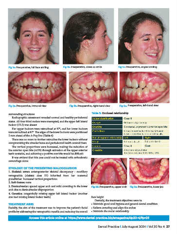
Fig 1a: Preoperative, full face smiling   Fig 1b: Preoperative, close-up smile   Fig 1c: Preoperative, angled smiling


            Fig 2a: Preoperative, intraoral view    Fig 2b: Preoperative, right-hand view   Fig 2c: Preoperative, left-hand view


           surrounding structures.                                 Table 5: Occlusal relationship
              Radiographic assessment revealed normal and healthy periodontal
           status. All four third molars were unerupted, and the upper left lateral
           incisor (UL2) was absent.
              Her upper incisors were retroclined at 97°, and her lower incisors
           were retroclined at 87°. The edges of the lower incisors were positioned
           5 mm ahead of the A-Pog line (Table 6).
              There was no room to further retrocline the lower incisors without
           compromising the alveolar bone and periodontal health around them.
              The vertical proportions were increased, making the reduction of
           the anterior open bite (AOB) through extrusion of the upper anterior
           teeth unstable, and achieving a positive overbite would be difficult.
              It was evident that this case could not be treated with orthodontic
           camouflage alone.

           ETIOLOGY OF THE PRESENTING MALOCCLUSION
           1. Skeletal: severe anteroposterior skeletal discrepancy - maxillary
           retrognathia (skeletal class III) inherited from her maternal
           grandfather. Increased vertical proportions
           2. Soft tissues: none
           3. Dentoalveolar: spaced upper arch and mild crowding in the lower   Fig 2d: Preoperative, upper arch  Fig 2e: Preoperative, lower jaw
           arch due to dentoalveolar disproportion
           4. Genetics:  congenitally missing upper left lateral incisor (mother
           also had missing lateral incisor teeth).                face height.
                                                                     Dentally, the treatment objectives were to:
           TREATMENT AIMS                                          • Maintain good oral hygiene and general dental condition
           Facially, the aim of the treatment was to improve the patient’s facial   • Relieve crowding and align the arches
           profile by addressing the retrognathic maxilla and reducing the overall   • Maintain the molar relationship

                         Access this article online at https://www.dental-practice.biz/emagazine/dp20-4/#p=26

                                                                       Dental Practice I July-August 2024 I Vol 20 No 4  27