Page 29 - DP Vol 20 No 4 HR
P. 29

upper dental midline to the right.
                                                                               A bilateral sagittal split osteotomy was performed
                                                                             using the Hunsuck and Dalpont modifications, with a
                                                                             setback.
                                                                               Finally, a genioplasty was carried out to balance the
                                                                             facial profile.

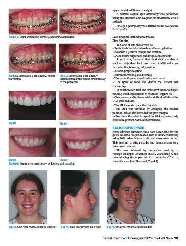
           Fig 5a-b: Eight weeks post-surgery, centerline correction         Post-Surgical Orthodontic Phase:
                                                                             Five Months
                                                                               The aims of this phase were to:
                                                                             • Settle the bite and achieve buccal interdigitation
                                                                             • Establish a positive overjet and overbite
                                                                             • Make minor alignment and torque adjustments
                                                                               At each visit, I ensured that the skeletal and dento-
                                                                             occlusal  objectives  had  been  met.  Additionally,  we
                                                                             recorded the following information:
                                                                             • Altered proprioception
           Fig 5c: Eight weeks post surgery, canine   Fig 5d: Eight weeks post surgery,   • Extraoral swelling and bruising
           correction                       lateralisation of the canine and intrusion   • The patient’s general well-being and mood
                                            of the premolar                  • The types of food and drinks the patient was
                                                                             consuming
                                                                               In collaboration with the restorative team, we began
                                                                             making small adjustments to the teeth (Figure 5):
                                                                             • Over several visits, the mesial and distal widths of the
                                                                             UL3 were reduced
                                                                             • The UL3 was also debulked buccally
                                                                             • The UL4 was intruded by changing the bracket
                                                                             position, which also intruded the gum margin
                                                                             • Over time, the palatal cusp of the UL4 was selectively
                                                                             ground to prevent occlusal interferences
           Fig 6a                           Fig 6b
                                                                             RESTORATIVE PHASE
                                                                             After allowing sufficient time post-debonding for the
                                                                             gums to settle, we proceeded with at-home whitening
                                                                             using 10% carbamide peroxide over a two-week period.
                                                                             This method is safe, reliable, and demonstrates very
                                                                             slow color rebound.
                                                                               This was followed by restorative masking to
                                                                             reshape the upper left canine (UL3), lateralizing it, and
                                                                             camouflaging  the  upper  left  first  premolar (UL4)  to
           Fig 6c                           Fig 6d                           resemble a canine (Figures 6, 7 and 8).
           Fig 6a-d: Restorative treatment – whitening and bonding


           Fig 7a: One-year review, full face smiling  Fig 7b: One-year review, side view    Fig 7c: One-year review, angled smiling


                                                                       Dental Practice I July-August 2024 I Vol 20 No 4  29
   24   25   26   27   28   29   30   31   32   33   34