
Page 50 - DP Vol 20 No 4 HR
P. 50
IMPLANT DENTISTRY SECTION
RESTORING FRACTURED MAXILLARY ANTERIOR
TOOTH BY EARLY LOADING OF SINGLE
TRABECULAR IMPLANT WITH A CHAIRSIDE
CROWN PLACEMENT ON TI-BASE ABUTMENT
AFTER 15 DAYS: A CASE REPORT
Gaurav Gupta, D. K. Gupta, Richa Gupta, Neelja Gupta
INTRODUCTION
Dental implants have been used in clinical practice for nearly
three decades and are widely accepted as a viable treatment
option for edentulous patients. Today, with advancements in
[1]
implant surface treatments, osseointegration is regarded as a
predictable biological process. However, while osseointegration
is crucial, it does not necessarily ensure patient satisfaction,
particularly in the aesthetic region. The success of dental
[2]
implant treatments hinges on both functional and aesthetic
outcomes. Smith and Zarb emphasized this by asserting that a
successful implant must not only function well but also meet
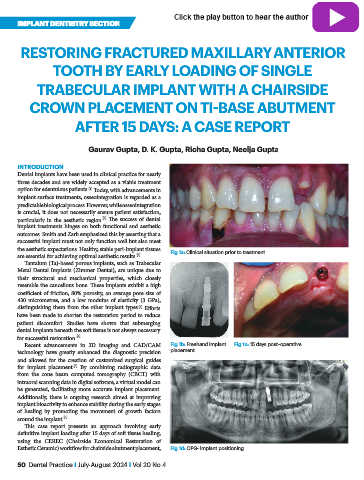
the aesthetic expectations. Healthy, stable peri-implant tissues Fig 1a: Clinical situation prior to treatment
are essential for achieving optimal aesthetic results. [2]
Tantalum (Ta)-based porous implants, such as Trabecular
Metal Dental Implants (Zimmer Dental), are unique due to
their structural and mechanical properties, which closely
resemble the cancellous bone. These implants exhibit a high
coefficient of friction, 80% porosity, an average pore size of
430 micrometres, and a low modulus of elasticity (3 GPa),
distinguishing them from the other implant types. Efforts
[3]
have been made to shorten the restoration period to reduce
patient discomfort. Studies have shown that submerging
dental implants beneath the soft tissue is not always necessary
for successful restoration. [4]
Recent advancements in 3D imaging and CAD/CAM Fig 1b: Freehand implant Fig 1c: 15 days post–operative
technology have greatly enhanced the diagnostic precision placement
and allowed for the creation of customized surgical guides
for implant placement. By combining radiographic data
[5]
from the cone beam computed tomography (CBCT) with
intraoral scanning data in digital software, a virtual model can
be generated, facilitating more accurate implant placement.
Additionally, there is ongoing research aimed at improving
implant bioactivity to enhance stability during the early stages
of healing by promoting the movement of growth factors
around the implant. [6]
This case report presents an approach involving early
definitive implant loading after 15 days of soft tissue healing,
using the CEREC (Chairside Economical Restoration of
Esthetic Ceramic) workflow for chairside abutment placement, Fig 1d: OPG- Implant positioning
50 Dental Practice I July-August 2024 I Vol 20 No 4