
Page 51 - DP Vol 20 No 4 HR
P. 51
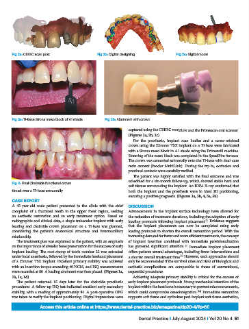
Fig 2a: CEREC scan post                 Fig 2b: Digital designing            Fig 2c: Digital model


            Fig 3a: Ti-Base Sirona meso block of A1 shade  Fig 3b: Abutment with crown

                                                                   captured using the CEREC workflow and the Primescan oral scanner.
                                                                   (Figures 2a, 2b, 2c)
                                                                     For the prosthesis, implant scan bodies and a screw-retained
                                                                   crown using the Zimmer TSX implant on a Ti-base were fabricated
                                                                   with a Sirona meso block in A1 shade using the Primemill machine.
                                                                   Sintering of the meso block was completed in the SpeedFire furnace.
                                                                   The crown was cemented extraorally onto the Ti-base with dual-cure
                                                                   resin cement (Ivoclar MultiLink). During the try-in, occlusion and
                                                                   proximal contacts were carefully verified.
                                                                     The patient was highly satisfied with the final outcome and was
                                                                   scheduled for a six-month follow-up, which showed stable hard and
            Fig 4: Final Chairside functional crown
                                                                   soft tissues surrounding the implant. An IOPA X-ray confirmed that
           timed over a Ti-base extraorally.                       both the implant and the prosthesis were in ideal 3D positioning,
                                                                   ensuring a positive prognosis. (Figures 3a, 3b, 4, 5a, 5b)
           CASE REPORT
           A  45-year-old  male  patient  presented  to  the  clinic  with  the  chief   DISCUSSION
           complaint of a fractured tooth in the upper front region, seeking   Advancements in the implant surface technology have allowed for
           an aesthetic restoration and an early treatment option. Based on   the reduction of treatment durations, including the adoption of early
           radiographic and clinical data, a single trabecular implant with early   loading protocols following implant placement.  Evidence suggests
                                                                                                       [7]
           loading  and chairside  crown placement on a  Ti-base  was planned,   that the implant placements can now be completed using early
           considering the patient’s anatomical structure and intermaxillary   loading protocols to shorten the overall restoration period. With the
           relationship.                                           increasing demand for faster and more efficient treatments, the concept
              The treatment plan was explained to the patient, with an emphasis   of implant insertion combined with immediate provisionalization
           on the importance of alveolar bone preservation for the success of early   has garnered significant attention.  Immediate implant placement
                                                                                             [8]
           implant loading. The root stump of tooth number 22 was extracted   offers patients several advantages, including fewer interventions and
           under local anesthesia, followed by the immediate freehand placement   a shorter overall treatment time.  However, such approaches should
                                                                                           [9]
           of a Zimmer TSX implant. Excellent primary stability was achieved   only be recommended if the survival rates and risks of biological and
           with an insertion torque exceeding 40 NCM, and ISQ measurements   aesthetic complications are comparable to those of conventional,
           were recorded at 80. A healing abutment was then placed. (Figures 1a,   sequential procedures.
           1b, 1c, 1d)                                               Achieving adequate primary stability is critical for the success of
              The patient returned 15 days later for the chairside prosthetic   early implant placement protocols. Strong mechanical retention of the
           procedures. A follow-up ISQ test indicated excellent early secondary   implant within the host bone is necessary to prevent micromovements,
           stability, with a reading of approximately 84. A post-operative OPG   which could compromise osseointegration.  Immediate restoration
                                                                                                    [10]
           was taken to verify the implant positioning. Digital impressions were   supports soft tissue and optimizes peri-implant soft tissue aesthetics,
                         Access this article online at https://www.dental-practice.biz/emagazine/dp20-4/#p=50

                                                                       Dental Practice I July-August 2024 I Vol 20 No 4  51