Page 36 - DP Vol 21 No1_Neat
P. 36

PEDIATRICS


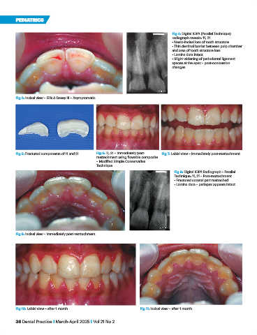
                                                                                      Fig 4: Digital IOPA (Parallel Technique)
                                                                                      radiograph reveals: 11, 21
                                                                                      • Mesio-incisal loss of tooth structure
                                                                                      • Thin dentinal barrier between pulp chamber
                                                                                      and area of tooth structure loss
                                                                                      • Lamina dura intact
                                                                                      • Slight widening of periodontal ligament
                                                                                      spaces at the apex – post-concussion
                                                                                      changes


           Fig 3: Incisal view – Ellis & Davey III – Asymptomatic.


           Fig 5: Fractured components of 11 and 21.  Fig 6: 11, 21 – Immediately post-  Fig 7: Labial view – immediately post-reattachment.
                                                  reattachment using flowable composite
                                                  – Modified Simple Conservative
                                                  Technique.
                                                                                       Fig 9: Digital IOPA Radiograph – Parallel
                                                                                       Technique: 11, 21 – Post-reattachment
                                                                                       • Fractured coronal part reattached
                                                                                       • Lamina dura – periapex appears intact


           Fig 8: Incisal view – immediately post-reattachment.


           Fig 10: Labial view – after 1 month.                       Fig 11: Incisal view – after 1 month.

           36 Dental Practice I March-April 2025 I Vol 21 No 2
   31   32   33   34   35   36   37   38   39   40   41