
Page 35 - DP Vol 21 No1_Neat
P. 35
modifying it to a V-shaped chamfer technique along 7. Vertical Groove
the fracture line. The chamfer is then restored with Two vertical grooves, 2mm in depth and width, are
composites. prepared in the labial surfaces after reattachment.
6. Overcontour Fiber-reinforced composite posts are placed in the
A groove is made along the fracture line – inciso- grooves and restored with composites.
apically – after reattachment. The groove is filled with Some studies have also indicated that the outcome
composite. Studies have reported the highest strength of reattachment is more technique-oriented than
with this technique compared to others. dependent on the type of material used.
Disadvantage: Discoloration of the composite over time.
The authors, prefer the Simple Conservative technique or its modification – The flowable composite is slightly overlapped over
the fracture margins after re-attachment and the same is subsequently finished well with spiral finishing burs. This also follows
Minimally Invasive Dentistry (M.I.D.) concepts. Moreover, the deeper understanding today, of concepts of post etch effects,
micro-tag formation, bonding agent infiltration into these micro-tags at the composite – tooth interface, hybrid layer formation,
greater adaptability of flowable composites to the tooth surfaces, higher strengths of nano- cluster flowable composite due
to increased filler content as well as higher aesthetic effects further strengthens the application of this technique over the
others. Another modification advocated in endodontically treated teeth: remove gutta percha from the pulp chamber and place
composites, which further aid retention of the fractured components.
Studies have shown that 34.2% of traumatized teeth develop pulpal Therefore, this necessitates regular clinical and radiological follow-
necrosis, necessitating endodontic treatment to prevent infection and/ ups in both the short and long term. Newer clinical interventions
or resorption to preserve the tooth. may need to be planned and applied if such changes are observed in
Post-reattachment, some pathological changes may occur over a subsequent follow-ups.
period of time: This prospective case highlights a positive outcome even after the
1. Fragment de-attachment patient stopped coming for review, and the sequelae could still be
2. Aesthetic concerns: Discolouration – this is the result of oxidation treated with a profound outcome even after 12 years—thus highlighting
of the haem part of the blood that has entered the dentinal tubules the advantage of seeking immediate care post-trauma. In literature, it
post-trauma. The extent of discolouration depends on the amount of is rare to see 10-year-plus follow-ups of reattachment protocols. This
blood ingress into the dentinal tubules at the time of trauma. case presents such an opportunity to share the outcome.
3. Intra-oral sinus – indicating pulpal necrosis.
4. Radiographically: CASE REPORT
a. Periapical radiolucent changes – these should be differentiated A 10-year-old girl fell down and traumatized her maxillary anterior
from post-concussion changes. teeth while playing at home. The girl’s family was aware enough to
b. Root resorption – internal as well as external. collect the fractured tooth fragments and give us a call. They were
c. Pulp canal calcifications (calcific metamorphosis) and subsequent informed about the need to place the fractured parts in cold water or
obliterations. cold milk and to reach the clinic as quickly as possible from a distance
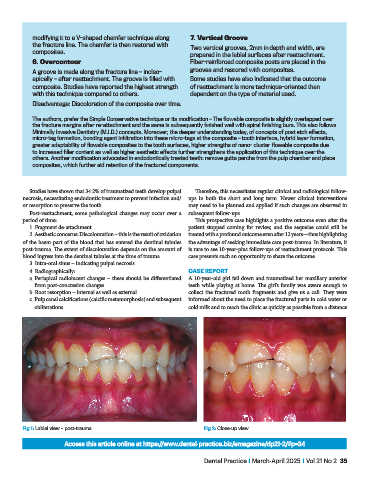
Fig 1: Labial view – post-trauma. Fig 2: Close-up view.
Access this article online at https://www.dental-practice.biz/emagazine/dp21-2/#p=34
Dental Practice I March-April 2025 I Vol 21 No 2 35