Page 37 - DP Vol 21 No1_Neat
P. 37

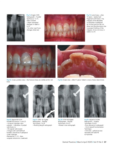
Fig 12: Digital IOPA                                             Fig 13: Labial view – after
                                 Radiograph – Parallel                                            12 years – reattached
                                 Technique: 11, 21 –                                              fragments still intact. The
                                 After 1 month                                                    margins of the flowable
                                 • Slight periapical                                              composites overlapped
                                 changes in relation                                              at the fracture line do not
                                 to 21                                                            show any discolouration due
                                 • No other significant                                           to proper finishing of the
                                 changes observed                                                 margins. Intra-oral sinus in
                                                                                                  relation to 21.


           Fig 14: Close-up labial view – the fracture lines are visible as thin red   Fig 15: Incisal view – after 12 years! Patient is now a heavy tea drinker.
           lines.


           Fig 16: Digital HD IOPA –    Fig 17: IOPA HD Digital    Fig 18: IOPA HD Digital     Fig 19: Digital HD IOPA
           Parallel Technique: 11 and 21  Radiograph – Parallel    Radiograph – Parallel       Radiograph – Parallel
           • Sclerotic changes intra-   Technique: 11 & 21         Technique: 11 & 21          Technique: 11 & 21
           pulpally – indicative of pulpal   • Working length radiograph  • Master cone radiograph  • Post-obturation radiograph
           calcifications                                                                      – Cold Lateral Condensation
           • Periapical radiolucent                                                            Technique
           changes with well-defined                                                           • Clinically – palatal access
           borders indicative of periapical                                                    restored with bulk-fill
           cystic changes                                                                      composites
           Planned for: 11, 21 – Non-
           surgical endodontic treatment
                                                                        Dental Practice I March-April 2025 I Vol 21 No 2  37
   32   33   34   35   36   37   38   39   40   41   42