
Page 16 - Dental Practice August 2022
P. 16
16-18-Anand Narvekar-Q8:18-22-Lanka Mahesh.qxd 8/17/2022 6:15 PM Page 1
prosthodontic section
STORY OF TWO ADJACENT PREMOLARS
ANAND NARVEKAR
CASE HISTORY
A patient was referred to the dental office to restore fractured tooth
no. 44 along with making new crowns on 44 and 45. History of joint
Zirconia crowns from which some part of tooth got fractured from
the tooth 44.
DIAGNOSIS
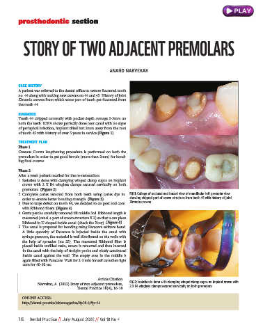
Tooth 44 chipped coronally with pocket depth average 2-3mm on
both the teeth. IOPA shows perfectly done root canal with no signs
of periapical infection, implant tilted but 2mm away from the root
of tooth 45 with history of over 5 years in service (Figure 1).
TREATMENT PLAN
Phase 1
Osseous Crown lengthening procedure is performed on both the
premolars in order to get good ferrule (more than 2mm) for bond-
ing final crowns.
Phase 2
After a week patient recalled for the re-restoration:
1. Isolation is done with clamping winged clamp supra on implant
crown with 2 X B4 wingless clamps secured cervically on both
premolars. (Figure 2)
2. Complete caries removal from both teeth using caries dye in FIG 1: Collage of occlusal and buccal view of mandibular left premolar view
order to ensure better bonding strength. (Figure 3) showing chipped part of crown structure from tooth 44 with history of joint
3. Due to large defect on tooth 44, we decided to do post and core Zirconia crowns
with Ribbond fibers. (Figure 4)
4. Gutta percha carefully removed till middle 3rd. Ribbond length is
measured (canal + part of crown structure X 2) so that u can place
Ribbond in U shaped inside canal (check the Xray). (Figure 4)
5. The canal is prepared for bonding using Paracore selfcure bond.
A little quantity of Paracore is injected inside the canal with
syringe pressure, the material is well distributed on the walls with
the help of spreader (no 25). The measured Ribbond fiber is
placed inside unfilled resin, excess is removed and then inserted
in the canal with the help of straight probe and nicely condensed
inside canal against the wall. The empty area in the middle is
again filled with Paracore. Wait for 2-3 min for self cure then light
cure for 40-60 sec.
Article Citation
Narvekar, A. (2022) Story of two adjacent premolars, FIG 2: Isolation is done with clamping winged clamp supra on implant crown with
Dental Practice 18(4), 16-18 2 X B4 wingless clamps secured cervically on both premolars
ONLINE ACCESS:
http://dental-practice.biz/emagazine/dp18-4/#p=16
16 Dental Practice // July-August 2022 // Vol 18 No 4