
Page 17 - Dental Practice August 2022
P. 17
16-18-Anand Narvekar-Q8:18-22-Lanka Mahesh.qxd 8/17/2022 6:15 PM Page 2
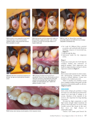
FIG 3: Complete caries removal from both teeth FIG 4: Gutta percha carefully removed till middle 3rd FIG 5: For tooth 45, Ribbond fibers encircled
using caries dye in order to ensure better Ribbond length is measured (canal + part of crown around the wall and build with Shofu low shrinkage
bonding strength, Canal prepared for ribbond structure X 2) so that you can place ribbond in U composite in incremental way.
fibers placement as a post shaped inside canal
6. For tooth 45, Ribbond fibers encircled
around the wall and build with Shofu low
shrinkage composite in incremental way.
(Figure 5)
7. Prep through the dam.
8. Impression made for the temporary
crown.
Phase 3
Temporary crowns given for 30-45 days for
complete healing and settlement of
gingival margins. Final impression was
made by using retraction cord and A-silicon
impression material. (Figure 7)
Phase 4
Both crowns made separate in order to main-
FIG 6: Both the teeth restored and ready for prep FIG 7: Collage of occlusal and buccal view of tain self-cleansing mechanism. Material
through the dam and impression for temporary mandibular left premolar view showing placement of selected is Lithium disilicate. Crowns were
crown retraction cord before final impression along with bonded in strict isolation protocols.
good ferrule to bond the final crown along with Taken 6 months post bonding crowns
healthy surrounding tissues post 1 month after show complete healing of the gums with the
placing temporary crowns.
formation of interdental papilla on all sides.
(Figures 8 and 9)
CONCLUSION
Joint crowns always give problems in long
term due to no self-cleaning mechanism like
natural teeth and leads to leakage in the
crowns. Selection of the material of the
crown is also important for long-standing
restoration.
Zirconia has high compressive as well
high tensile strength which transforms all
stresses to the tooth and if the tooth is weak
(heavily restored like shown in this case)
then there are high chances of
catastrophic failure.
FIG 8: Occlusal view showing natural anatomy of both separate crowns. Osseous CLP creates good ferrule for
Dental Practice // July-August 2022 // Vol 18 No 4 17