
Page 44 - DP.qxd
P. 44
Implantology sectIon
Putty Assisted Sinus Augmentation:
The “Pasa Technique” Discussion
and A Descriptive Case
Lanka Mahesh, Glenn Mascarenhas, Sagrika Shukla, Tanvi Paliwal, Zara Dhawan
ABSTRACT
GBR with bone graft for sinus augmentation are well established
techniques in implant dentistry. Technique based on PASS
principle has predictable regeneration and wound healing after
implant placement. Implant design and its surface characteristics
also play a major role when sinus augmentation and implants are
placed simultaneously.
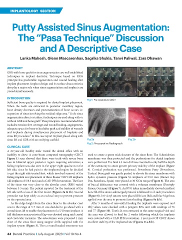
iNTRODUCTiON Fig 1: Pre operative CBCT
Sufficient bone quality is required for dental implant placement.
When the teeth are extracted in posterior maxillary region,
bone density decreases and pneumatization of bone occurs i.e.
1
expansion of sinus involving the residual ridge area. To do sinus
augmentation direct or indirect techniques are used along with or
2
without GBR and bone graft. Pass principle is recommended that
includes: tension free coverage and wound healing, angiogenesis,
adequate space for bone to heal after graft and stability of wounds
and implants during simultaneous placement of Implants and
3
sinus lift procedure. In this case report implant placed along with
sinus lift and GBR with an ossifying scaffold. Fig 2a Fig 2b
Fig 2: Preoperative Radiograph
CLiNiCAL CASE
A 62-year-old healthy male visited the dental office with an
inability to chew. A cone-beam computed tomography (CBCT used to create a green stick fracture of the sinus floor. The Schneiderian
Figure 1) scan showed that there were teeth with severe bone membrane was then protected and the perforations for dental implants
loss in bilateral upper posterior region requiring extraction; a were performed. The final 4.3 mm drill was inserted to only half the depth
failing implant was also seen in the upper right quadrant on the of the osteotomy to attain greater primary stability of the implant (Figure
left side, since he had pain in the implanted region he decided 4). Cortical perforation was performed. Powerbone Putty (Powerbone,
to get the right side treated first, which involved removal of the Turkey) Bone graft was gently pushed to elevate the sinus membrane with
failing implant and placement of three Bioner TOP DM implants hydro dynamic pressure (Figure 5). Implants of 5/10 mm (Bioner Top
(all implants of 5/8.5 mm) and a CAD PFM restoration. The floor Dm, Barcelona, Spain) were placed at 30 NCm torque (Figure 6). The area
of the sinus was very close to the alveolar crest. (RBH varied of buccal dehiscence was covered with a volumax membrane (Dentsply
between 3-5 mm). The patient reported for the treatment of the Sirona, Germany) (Figure 7). An RVG taken immediately showed excellent
left side with a loss of the first molar (Figures 2a & 2b). Second bone fill of the sinus a submerged protocol is followed in all such procedures
premolar was kept solely as an occlusal stop to prevent pressure (Figure 8). 3-0 vicryl sutures were placed (Ethicon J&J) and blue M gel was
on the operated area. applied over the area to promote faster healing (Figures 9a & b).
As the ridge height from the sinus floor to the alveolar crest After 5 months of uneventful healing the implants were exposed and
was in the range of 5-7 mm, it was decided to go ahead with a ISQ values were checked with a penguin RFA unit with readings of 76
crestal sinus lift to enable the placement of a 5/10 mm implant. A and 77 (Figure 10). Tooth 24 was extracted at the same surgical visit and
full thickness mucoperiosteal flap was elevated using mid crestal the area was allowed to heal for 2 weeks following which the implants
and crevicular incisions. The osteotomies were prepared 1 mm were restored with a CAD PFM restoration. 1 year post OP CBCT shows
short of the sinus floor using stopper drills supplied with the excellent stability of the implanted site (Figures 11a & b).
implant system (Figure 3). Then a round headed osteotome was
44 Dental Practice i July-August 2023 i Vol 19 No 4