
Page 20 - DP20-2.qxd
P. 20
implantology section
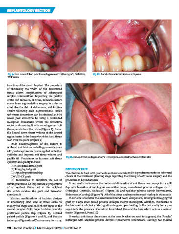
Fig 9: Non cross-linked porcine collagen matrix (Mucograft, Geistlich, Fig 10: Band of keratinized tissue at 3 years
Wolhusen
insertion of the dental implant. The procedure
of increasing the width of the keratinized
tissue allows simplification of subsequent
surgical interventions. Improving the quality
of the soft tissues is, at times, indicated before
major bone augmentation surgery in order to
minimize the risk of dehiscence, which often
occurs following such augmentations. Stable
soft-tissue dimensions can be obtained at 8-10
weeks post extraction by using a controlled
resorption biomaterial within the extraction
socket and covering it with an autogenous soft
tissue punch from the palate (Figure 1). Better
the bound down tissue volume at the crestal
region better is the longevity of the hard tissue
seen over the years. (Figure 2)
Once osseointegration of the fixture is
achieved and basic remodelling process is done
with, various protocols can be applied to further
optimize and improve soft tissue volume and
papilla fill. Procedures to increase soft tissue Fig 11: Cross-linked collagen matrix - Fibrogide, adapted to the recipient site
quantity and quality include:
(A) Connective tissue graft
(B) Free gingival graft DECISION TREE
(C) Apically positioned flap The clinician is flush with protocols and biomaterials and it is prudent to make an informed
(D) VIP-CT graft choice at the treatment planning stage regarding the timing of soft tissue surgery and the
(E) Biomaterials to substitute the use of procedure to be undertaken.
autologous tissue. Of importance is preparation If our goal is to increase the horizontal dimension of soft tissue, we can opt for a split
of an optimal tissue bed at the recipient flap with insertion of autologous connective tissue, cross-linked porcine collagen matrix
site which receives the graft and thereafter (Fibrogide, Geistlich, Wolhusen) (Figure 11) and acellular porcine dermis (Novomatrix,
nourishes it. Biohorizons Camlog) (Figure 7). All of the above undergo submerged healing in the mouth.
Various modifications of incision designs If our aim is to better the keratinized bound down component, autologous free gingival
at uncovering exist and at times serve to graft or a non cross-linked porcine collagen matrix (Mucograft, Geistlich, Wolhusen) is
modify the shape and bulk of soft tissue at the the biomaterial of choice. Mucograft undergoes open healing in the oral cavity but a pre-
crestal complex. Split finger incision, laterally requisite is the presence of minimal keratinized tissue at the base which acts as a cellular
positioned pedicle flap (Figure 3), Rotated feeder. (Figures 8, 9 and 10)
palatal pedicle (Figures 4 and 5), and Poncho If vertical soft tissue dimensions at the crest is what we need to augment, the ‘Poncho’
technique (Figure 6 and 7) are among the many. technique with acellular porcine dermis (Novomatrix, Biohorizons Camlog) has showed
20 Dental Practice I March-April 2024 I Vol 20 No 2