
Page 40 - DP Vol 20 No 4 HR
P. 40
PEDIATRIC DENTISTRY SECTION
ADVANCED RESTORATION OF PRIMARY
ANTERIOR TEETH WITH CAD/CAM
TECHNOLOGY: A CASE REPORT
Joies Susan Varghese, PC Chayanika, Smita Kumari
INTRODUCTION
Primary teeth are crucial elements of a child’s dentition, and their
importance is increasingly recognized. Preserving their functional
and aesthetic qualities, supporting normal speech development, and
maintaining adequate space for the eruption of permanent teeth are
key reasons why primary teeth are treated with utmost care.¹
CAD/CAM (Computer-Aided Design and Computer-Aided
Manufacturing) systems have been used in dentistry since the mid-
1980s and have gained popularity due to their wide application in
various fields, including prosthodontics, crown and bridge work,
implantology, orthodontics, and pediatric dentistry.² This technology
provides aesthetic restorations that are both durable and precise. The
exceptional mechanical properties of these materials make CAD/
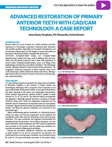
CAM technology a reliable method for dental restorations, offering a Fig 1: Pre-treatment view
high survival rate and minimal incidence of fractures, ensuring long-
term clinical durability.³
CASE REPORT
A 5-year-old female patient presented to the Department of Pediatric
& Preventive Dentistry at the Institute of Dental Studies and
Technologies, Modinagar, with a complaint of food impaction in the
upper anterior teeth for the past two weeks. A thorough medical history
was taken, followed by the clinical and radiographic examinations,
which revealed deep proximal dentinal caries in relation to teeth
numbers 54, 51, 61, 64, and 84. The child was highly uncooperative
and displayed a “definitely negative” behaviour according to Frankl’s
behaviour rating scale. Given her behavioural assessment and the
extensive need for treatment, it was decided to treat her under general
anaesthesia. Signed informed consent was obtained from the patient’s Fig 2: After teeth preparation
parents, and fitness for general anaesthesia was confirmed by the
anaesthesiologist.
Before administering general anaesthesia, an orthopantomogram
(OPG) was taken, and intraoral impressions of the maxillary and
mandibular arches were made for fabricating a band and loop space
maintainer. The space maintainer was prepared conventionally and
cemented during the procedure. The maxillary impression was also
used to fabricate a custom impression tray. NPO (nil per os) guidelines
were provided to the parents, and the patient was scheduled for
operative intervention under general anaesthesia.
During the procedure, endodontic treatment was performed
on teeth numbers 51, 61, and 54, and tooth 84 was extracted. The
prefabricated band and loop space maintainer was cemented in place
for tooth 84. For post-endodontic restoration of the anterior teeth, Fig 3: Custom-made impression tray
40 Dental Practice I July-August 2024 I Vol 20 No 4