
Page 35 - DP Vol 20 No 4 HR
P. 35
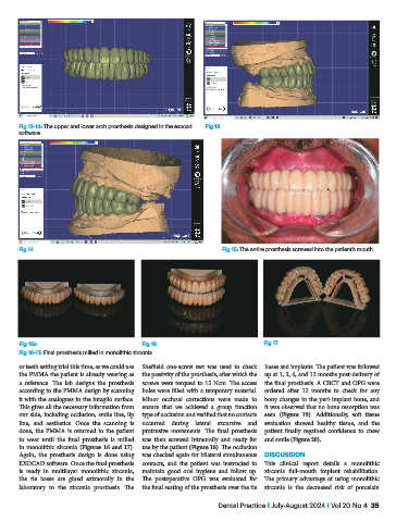
Fig 12-14: The upper and lower arch prosthesis designed in the exocad   Fig 13
           software


           Fig 14                                                       Fig 15: The entire prosthesis screwed into the patient’s mouth


           Fig 16a                              Fig 16                              Fig 17
           Fig 16-17: Final prosthesis milled in monolithic zirconia

           or teeth setting trial this time, as we could use   Sheffield one-screw test was used to check   bases and implants. The patient was followed
           the PMMA the patient is already wearing as   the passivity of the prosthesis, after which the   up at 1, 3, 6, and 12 months post-delivery of
           a  reference.  The  lab  designs  the  prosthesis   screws were torqued to 15 Ncm. The access   the final prosthesis. A CBCT and OPG were
           according to the PMMA design by scanning   holes were filled with a temporary material.   ordered after 12 months to check for any
           it with the analogues in the intaglio surface.   Minor occlusal corrections were made to   bony changes in the peri-implant bone, and
           This gives all the necessary information from   ensure that we achieved a group function   it was observed that no bone resorption was
           our side, including occlusion, smile line, lip   type of occlusion and verified that no contacts   seen  (Figure 19). Additionally, soft tissue
           line, and aesthetics. Once the scanning is   occurred during lateral excursive and   evaluation showed healthy tissue, and the
           done, the PMMA is returned to the patient   protrusive movements. The final prosthesis   patient finally regained confidence to chew
           to wear until the final prosthesis is milled   was then screwed intraorally and ready for   and smile (Figure 20).
           in monolithic zirconia (Figures 16 and 17).   use by the patient (Figure 18). The occlusion
           Again, the prosthesis design is done using   was checked again for bilateral simultaneous   DISCUSSION
           EXOCAD software. Once the final prosthesis   contacts, and the patient was instructed to   This clinical report details a monolithic
           is ready in multilayer monolithic zirconia,   maintain good oral hygiene and follow up.   zirconia full-mouth implant rehabilitation.
           the tie bases are glued extraorally in the   The postoperative OPG was evaluated for   The  primary  advantage  of using monolithic
           laboratory to the zirconia prosthesis. The   the final seating of the prosthesis over the tie   zirconia is the decreased risk of porcelain

                                                                       Dental Practice I July-August 2024 I Vol 20 No 4  35