
Page 57 - DP Vol 20 No 4 HR
P. 57
shape regardless of how much it is deflected, theoretically enhancing a horizontal growth pattern with FMA of 20°. Soft tissue analysis
treatment precision and efficiency. 13 revealed an obtuse nasolabial angle of 99° and a lip strain of 3mm
The treatment can be staged depending upon the severity of the (Figure 7b). The OPG findings revealed missing maxillary right and
malocclusion, as mentioned below: left third molars. (Figure 7a)
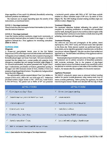
LEVEL 1- One stage treatment Treatment Objectives
These include the cases that require the least intervention by the doctor The treatment objectives included addressing the patient’s chief
over the course of treatment. complaint by correcting proclination of maxillary and mandibular
anterior teeth, closing the spaces in the maxillary anterior region while
LEVEL 2- Two stage treatment maintaining Class I canine and molar relation on both sides along with
Cases that involve auxiliary mechanics, staged tooth movements, or achieving a harmonious soft tissue profile.
more complex treatment plans are treated in two stages i.e. An initial
appliance followed by a final appliance for remaining corrections. Treatment Planning
(Refer to Table 2) The extra-oral and intra-oral photographs of the patient, lateral
cephalogram, OPG, CBCT scans, and intraoral scans were uploaded
CLINICAL CASE on the Brava site. Finite element analysis was performed based on
Diagnosis these details, and an algorithm generated a treatment plan on the Brius
A 24-year-old post-pubertal female came to the Out Patient case planner software (Figure 8). This plan was then cross-verified and
Department (OPD) of the Department of Orthodontics and Dentofacial approved by us. The fabrication and delivery of the appliance took two
Orthopedics of our institution, with the chief complaint of spacing and weeks to complete.
forwardly placed upper and lower front teeth. Extraoral examination Treatment planning included the extraction of the upper and lower
revealed that the patient had a convex profile with posterior facial first premolars and to achieve correction of bimaxillary protrusion
divergence, competent lips and average nasolabial angle (Figure 5). with moderate anchorage. Due to the presence of peg-shaped
On intraoral examination, the patient presented with Angle’s Class I maxillary lateral incisors and Bolton’s excess in the mandibular arch it
type 2 malocclusion, proclination of incisors, generalized spacing in was planned to maintain spaces on both sides of the maxillary lateral
the upper arch with increased overjet of 3mm, overbite of 3mm and incisors, for veneers after the completion of orthodontic treatment.
presence of peg shaped lateral incisors. The patient also had generalized
dental fluorosis. (Figure 6) Appliance Placement
The cephalometric analysis revealed skeletal Class I jaw relation on The BRIUS® orthodontic system uses an advanced indirect bonding
account of SNA of 84˚, SNB of 82˚ and ANB angle of 2˚. Patient had method for precise bracket placement, using custom-made trays to
proclined incisors (Upper incisor to NA= 34°/8 mm, Lower incisor ensure accurate positioning. Before placing the appliance, the teeth
to NB=30°/7mm). The patient had an IMPA of 99° and presented were thoroughly cleaned. Cheeks were retracted with the help of cheek
Table 2: Case selection for One stage or Two stage treatment
Dental Practice I July-August 2024 I Vol 20 No 4 57