
Page 58 - DP Vol 20 No 4 HR
P. 58
ORTHODONTIC SECTION
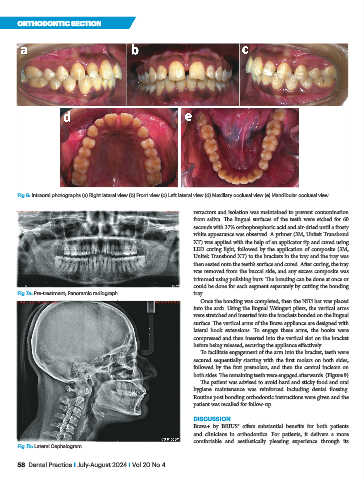
Fig 6: Intraoral photographs (a) Right lateral view (b) Front view (c) Left lateral view (d) Maxillary occlusal view (e) Mandibular occlusal view
retractors and isolation was maintained to prevent contamination
from saliva. The lingual surfaces of the teeth were etched for 60
seconds with 37% orthophosphoric acid and air-dried until a frosty
white appearance was observed. A primer (3M, Unitek Transbond
XT) was applied with the help of an applicator tip and cured using
LED curing light, followed by the application of composite (3M,
Unitek Transbond XT) to the brackets in the tray and the tray was
then seated onto the teeth’s surface and cured. After curing, the tray
was removed from the buccal side, and any excess composite was
trimmed using polishing burs. The bonding can be done at once or
could be done for each segment separately by cutting the bonding
Fig 7a: Pre-treatment; Panoramic radiograph tray.
Once the bonding was completed, then the NiTi bar was placed
into the arch. Using the lingual Weingart pliers, the vertical arms
were stretched and inserted into the brackets bonded on the lingual
surface. The vertical arms of the Brava appliance are designed with
lateral hook extensions. To engage these arms, the hooks were
compressed and then inserted into the vertical slot on the bracket
before being released, securing the appliance effectively.
To facilitate engagement of the arm into the bracket, teeth were
secured sequentially starting with the first molars on both sides,
followed by the first premolars, and then the central incisors on
both sides. The remaining teeth were engaged afterwards. (Figure 9)
The patient was advised to avoid hard and sticky food and oral
hygiene maintenance was reinforced including dental flossing.
Routine post bonding orthodontic instructions were given and the
patient was recalled for follow-up.
DISCUSSION
Brava+ by BRIUS® offers substantial benefits for both patients
and clinicians in orthodontics. For patients, it delivers a more
comfortable and aesthetically pleasing experience through its
Fig 7b: Lateral Cephalogram
58 Dental Practice I July-August 2024 I Vol 20 No 4